Copyright
©The Author(s) 2024.
World J Hepatol. Feb 27, 2024; 16(2): 279-285
Published online Feb 27, 2024. doi: 10.4254/wjh.v16.i2.279
Published online Feb 27, 2024. doi: 10.4254/wjh.v16.i2.279
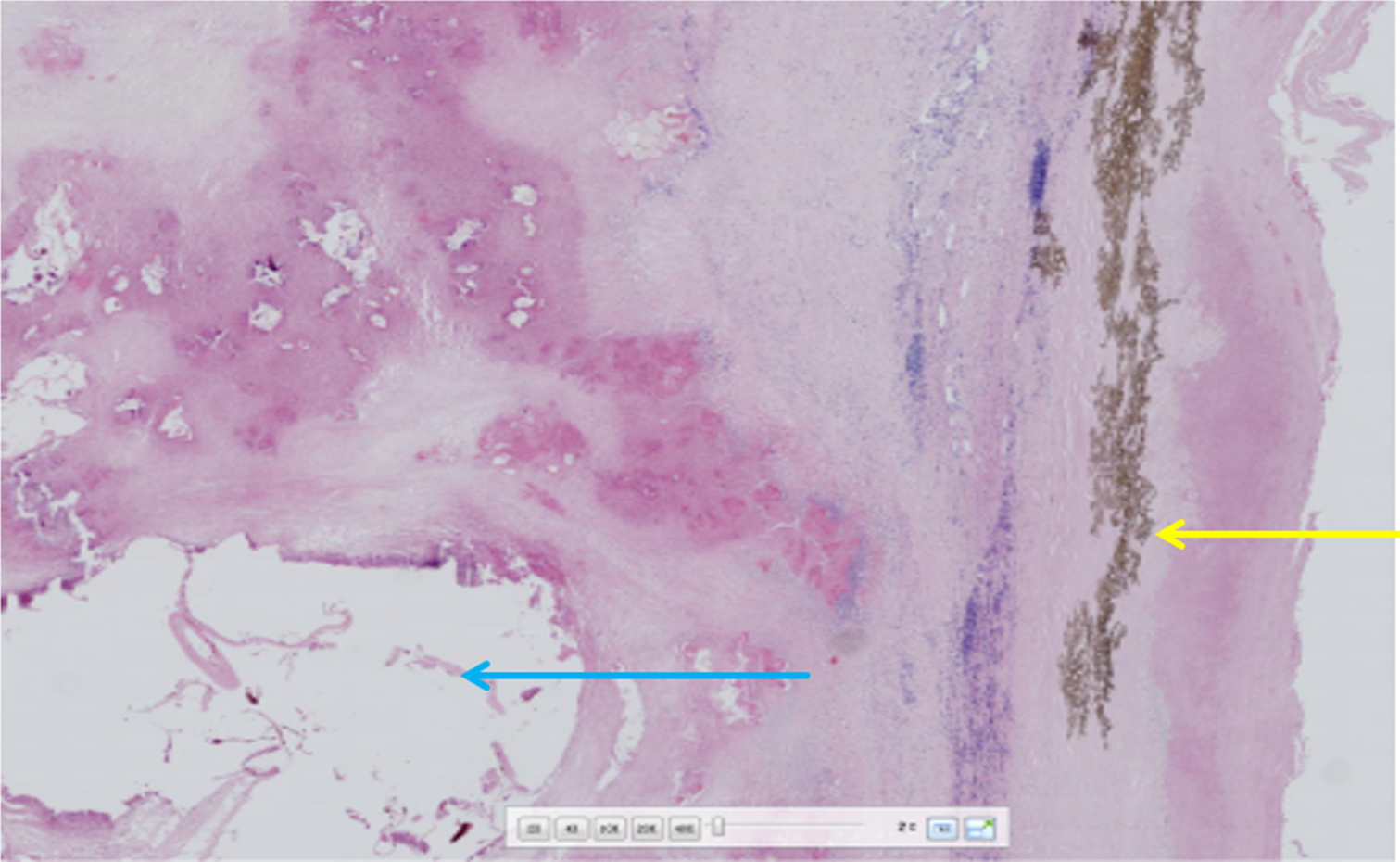
Figure 4
Postoperative pathology slides Histopathological examination by hemotoxylin-eosin staining (200 ×) fibrous connective tissue proliferation and inflammatory cell infiltration are seen around the blue arrow vesicles, forming nodules of varying sizes (alveolar echinococcosis) yellow arrow laminar-like structures are clearly visible (cystic echinococcosis).
- Citation: Wang MM, An XQ, Chai JP, Yang JY, A JD, A XR. Coinfection with hepatic cystic and alveolar echinococcosis with abdominal wall abscess and sinus tract formation: A case report. World J Hepatol 2024; 16(2): 279-285
- URL: https://www.wjgnet.com/1948-5182/full/v16/i2/279.htm
- DOI: https://dx.doi.org/10.4254/wjh.v16.i2.279


