Copyright
©The Author(s) 2024.
World J Hepatol. Dec 27, 2024; 16(12): 1505-1514
Published online Dec 27, 2024. doi: 10.4254/wjh.v16.i12.1505
Published online Dec 27, 2024. doi: 10.4254/wjh.v16.i12.1505
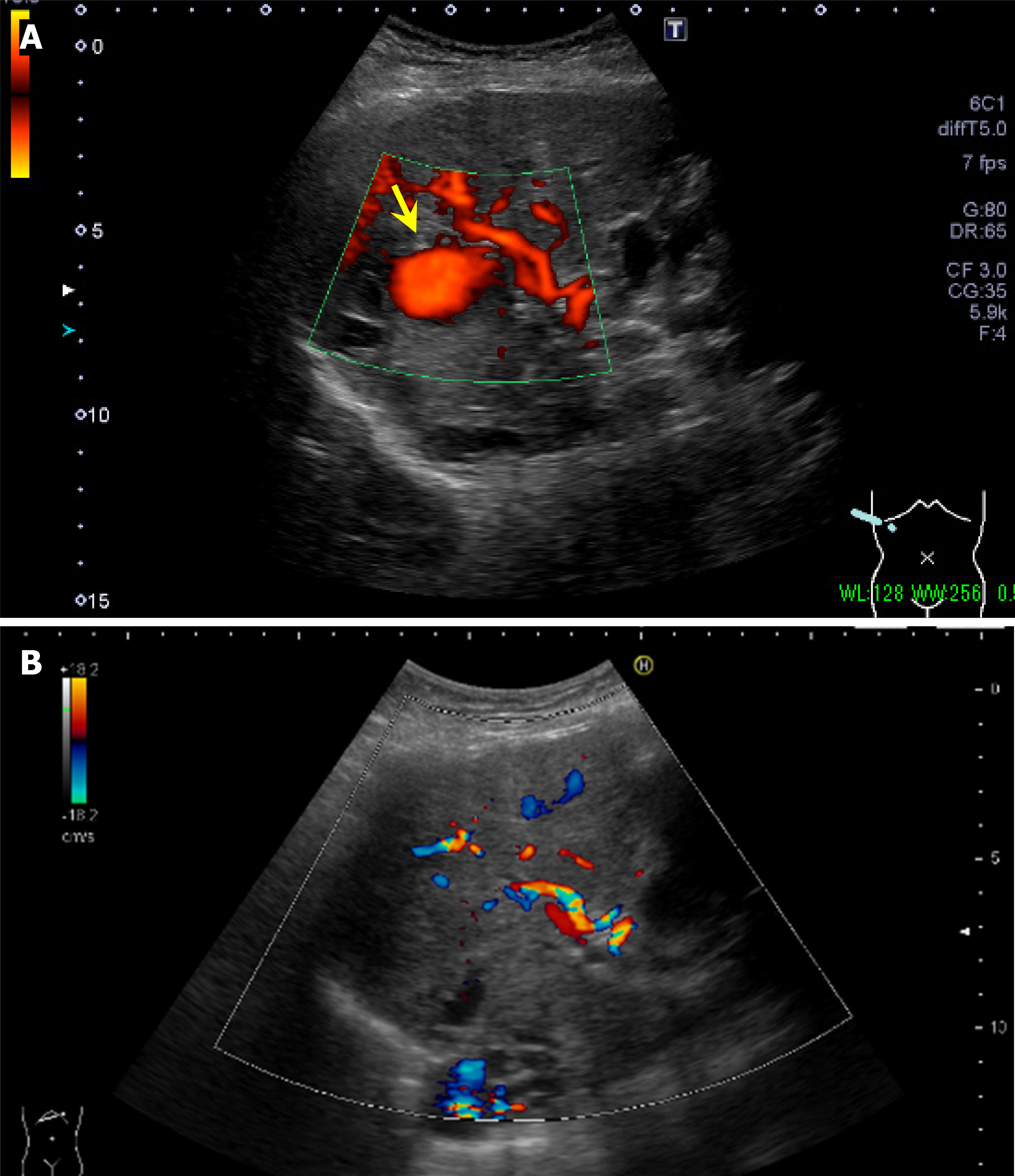
Figure 3 Abdominal ultrasonography.
A: Abdominal ultrasonography before aneurysm embolization. A mass lesion at hepatic S 7/8 showing pulsating blood flow (yellow arrow) was diagnosed as a hepatic artery aneurysm; B: Abdominal ultrasonography after aneurysm embolization. The intrahepatic artery aneurysm seen before aneurysm embolization had disappeared.
- Citation: Tamura H, Ozono Y, Uchida K, Uchiyama N, Hatada H, Ogawa S, Iwakiri H, Kawakami H. Multiple intrahepatic artery aneurysms during the treatment for IgG4-related sclerosing cholangitis: A case report. World J Hepatol 2024; 16(12): 1505-1514
- URL: https://www.wjgnet.com/1948-5182/full/v16/i12/1505.htm
- DOI: https://dx.doi.org/10.4254/wjh.v16.i12.1505


