Copyright
©The Author(s) 2024.
World J Hepatol. Dec 27, 2024; 16(12): 1505-1514
Published online Dec 27, 2024. doi: 10.4254/wjh.v16.i12.1505
Published online Dec 27, 2024. doi: 10.4254/wjh.v16.i12.1505
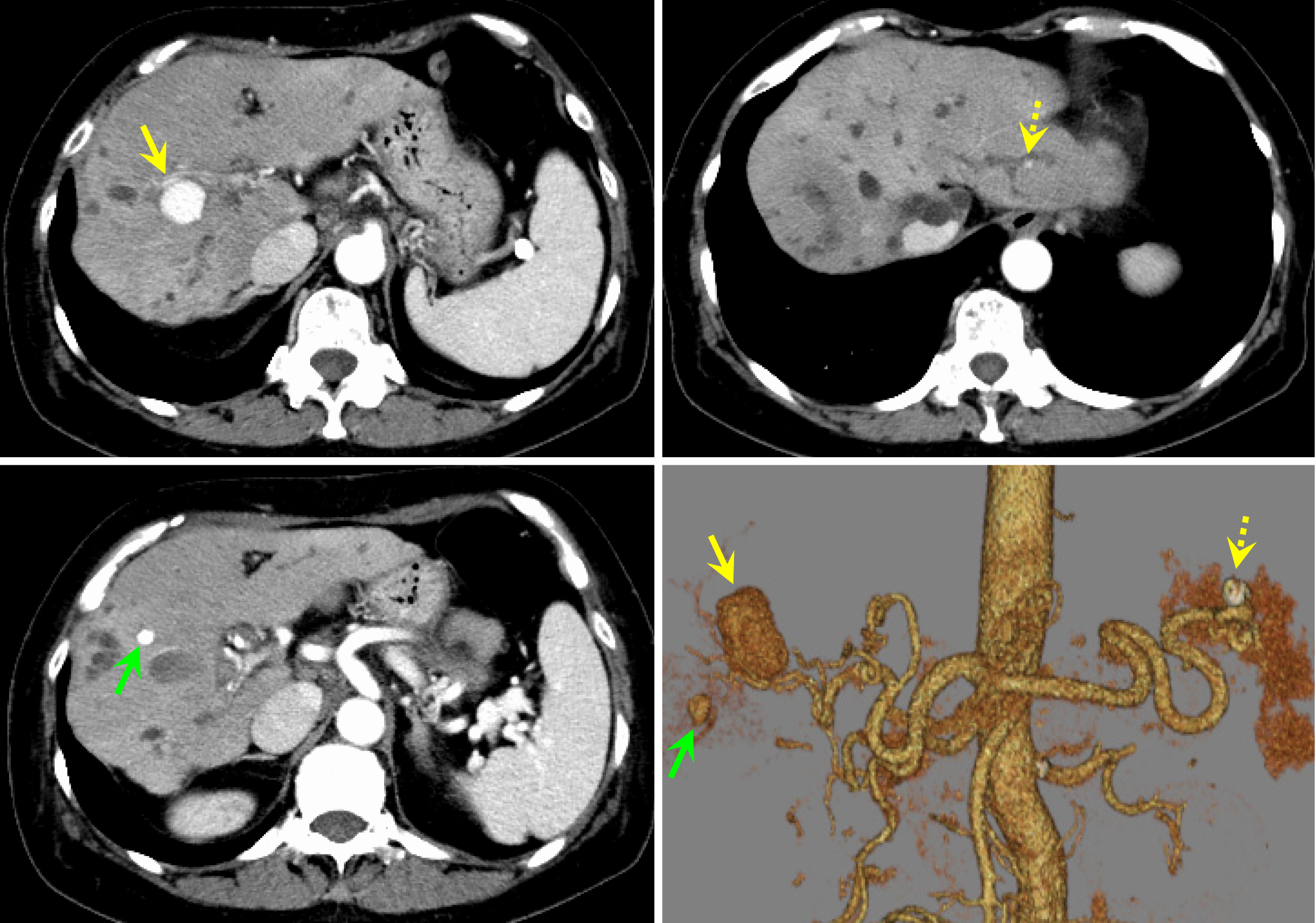
Figure 2 Contrast-enhanced computed tomography scan of the abdomen at diagnosis.
Intrahepatic artery aneurysms were suspected as nodular contrast-enhanced areas approximately 20 mm in size appeared in S 7/8 (yellow arrow), 7 mm in size in S 5 (green arrow), and 4 mm in size in the lateral segment (dotted yellow arrow).
- Citation: Tamura H, Ozono Y, Uchida K, Uchiyama N, Hatada H, Ogawa S, Iwakiri H, Kawakami H. Multiple intrahepatic artery aneurysms during the treatment for IgG4-related sclerosing cholangitis: A case report. World J Hepatol 2024; 16(12): 1505-1514
- URL: https://www.wjgnet.com/1948-5182/full/v16/i12/1505.htm
- DOI: https://dx.doi.org/10.4254/wjh.v16.i12.1505


