Copyright
©The Author(s) 2024.
World J Hepatol. Nov 27, 2024; 16(11): 1348-1355
Published online Nov 27, 2024. doi: 10.4254/wjh.v16.i11.1348
Published online Nov 27, 2024. doi: 10.4254/wjh.v16.i11.1348
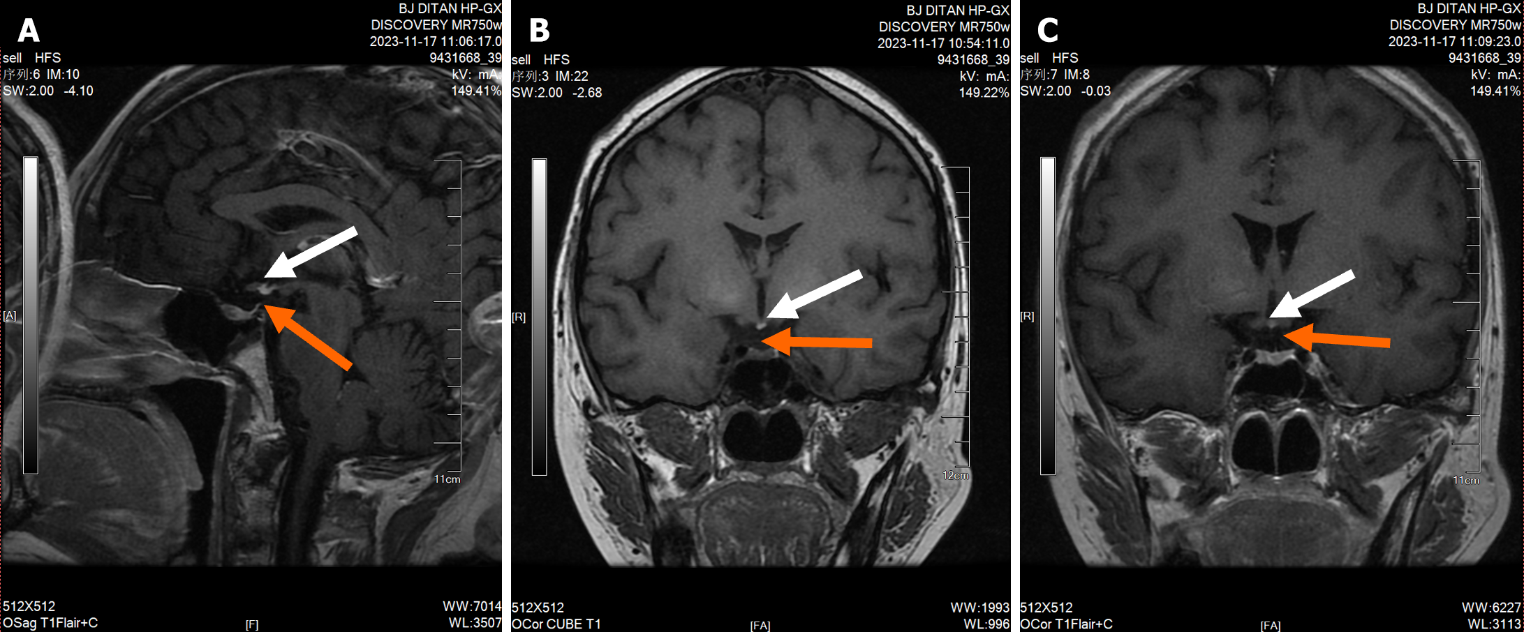
Figure 4 Cranial magnetic resonance imaging results.
Absence of pituitary stalk and ectopic posterior pituitary. The red arrow shows the position of the pituitary stalk, and the white arrow indicates the ectopic posterior pituitary. A: The mid-sagittal plane showed a normal pituitary volume, absence of visualization of the pituitary stalk, and loss of the normal hyperintensity of the posterior pituitary lobe. An ectopic high signal was observed at the infundibular recess; B and C: Coronal planes demonstrated the absence of visualization of the pituitary stalk and loss of the normal hyperintensity of the posterior pituitary lobe, with an ectopic high signal at the infundibular recess.
- Citation: Chang M, Wang SY, Zhang ZY, Hao HX, Li XG, Li JJ, Xie Y, Li MH. Pituitary stalk interruption syndrome complicated with liver cirrhosis: A case report. World J Hepatol 2024; 16(11): 1348-1355
- URL: https://www.wjgnet.com/1948-5182/full/v16/i11/1348.htm
- DOI: https://dx.doi.org/10.4254/wjh.v16.i11.1348


