Copyright
©The Author(s) 2023.
World J Hepatol. May 27, 2023; 15(5): 715-724
Published online May 27, 2023. doi: 10.4254/wjh.v15.i5.715
Published online May 27, 2023. doi: 10.4254/wjh.v15.i5.715
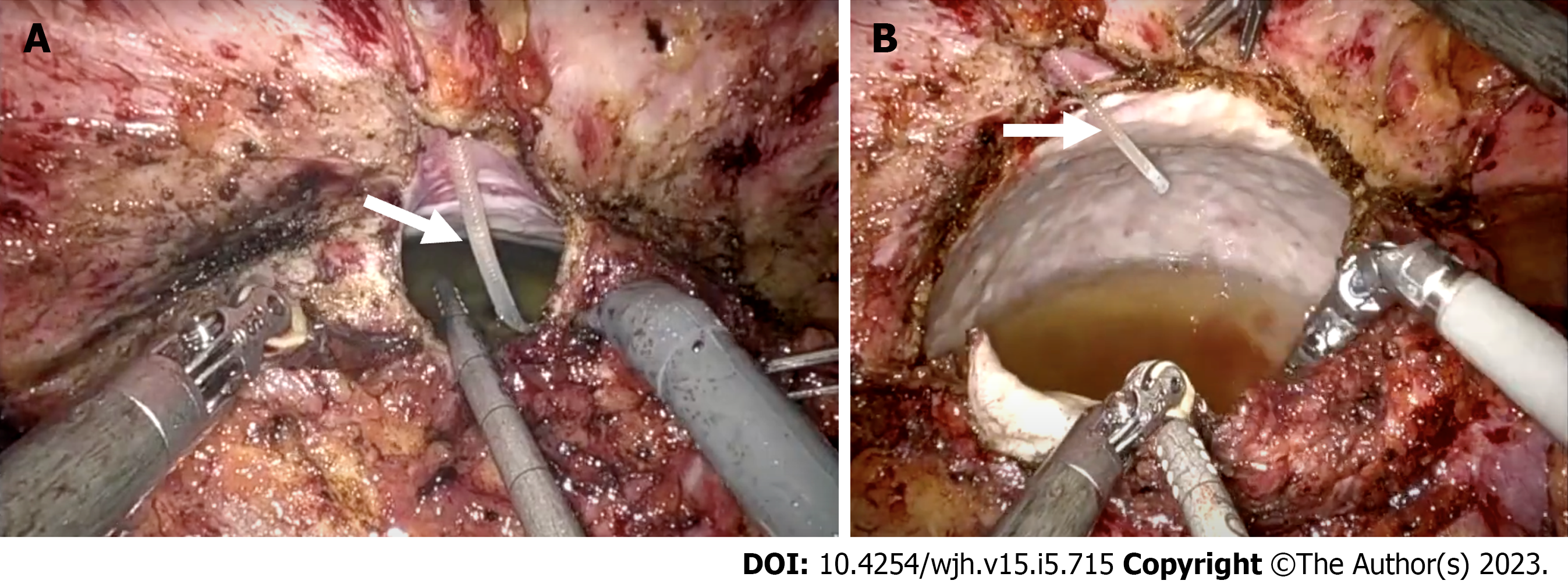
Figure 4 Intraoperative images of hepatic cerebrospinal fluid pseudocyst.
A: A large right hepatic lobe pseudocyst with tip of right ventriculoperitoneal shunt catheter within the cyst cavity (arrow); B: A large volume of cerebrospinal fluid can be seen within the cyst cavity (arrow).
- Citation: Yousaf MN, Naqvi HA, Kane S, Chaudhary FS, Hawksworth J, Nayar VV, Faust TW. Cerebrospinal fluid liver pseudocyst: A bizarre long-term complication of ventriculoperitoneal shunt: A case report. World J Hepatol 2023; 15(5): 715-724
- URL: https://www.wjgnet.com/1948-5182/full/v15/i5/715.htm
- DOI: https://dx.doi.org/10.4254/wjh.v15.i5.715


