Copyright
©The Author(s) 2023.
World J Hepatol. Apr 27, 2023; 15(4): 538-553
Published online Apr 27, 2023. doi: 10.4254/wjh.v15.i4.538
Published online Apr 27, 2023. doi: 10.4254/wjh.v15.i4.538
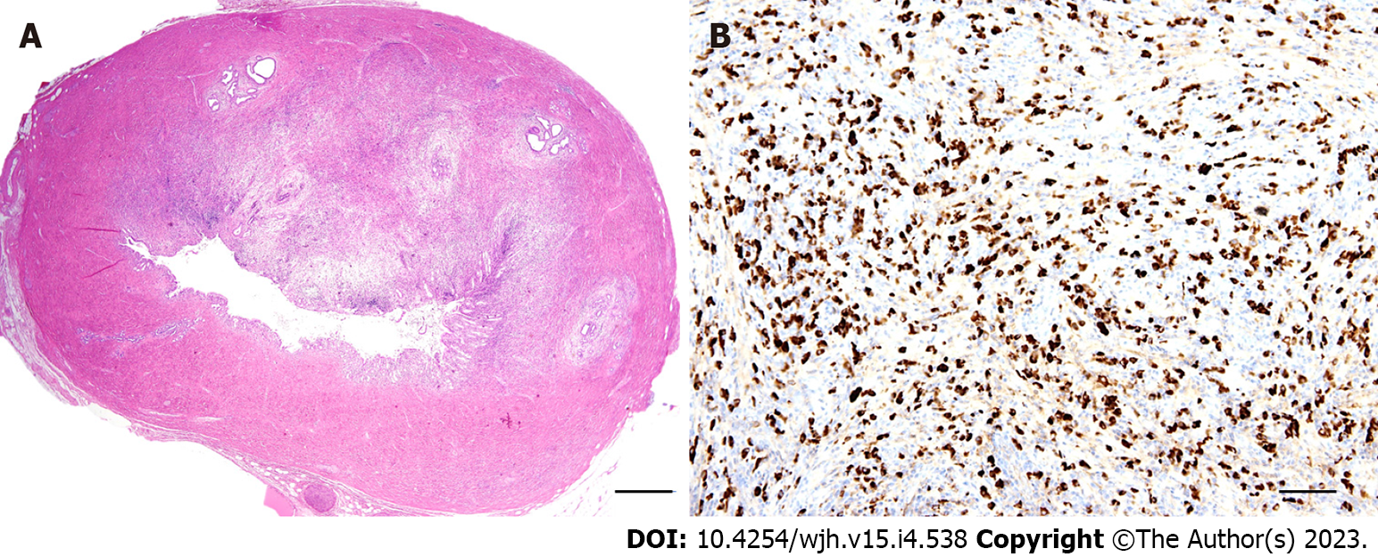
Figure 5 Morphology of IgG4-related sclerosing cholangiopathy.
A: Marked fibroinflammatory thickening of the bile duct wall; B: Increased numbers of IgG4-positive plasma cells. Haematoxylin and eosin (A), IgG4 immunohistochemistry (B). Bar corresponds to 1000 µm (A) and 100 µm (B).
- Citation: Sticova E, Fabian O. Morphological aspects of small-duct cholangiopathies: A minireview. World J Hepatol 2023; 15(4): 538-553
- URL: https://www.wjgnet.com/1948-5182/full/v15/i4/538.htm
- DOI: https://dx.doi.org/10.4254/wjh.v15.i4.538


