Copyright
©The Author(s) 2023.
World J Hepatol. Oct 27, 2023; 15(10): 1140-1152
Published online Oct 27, 2023. doi: 10.4254/wjh.v15.i10.1140
Published online Oct 27, 2023. doi: 10.4254/wjh.v15.i10.1140
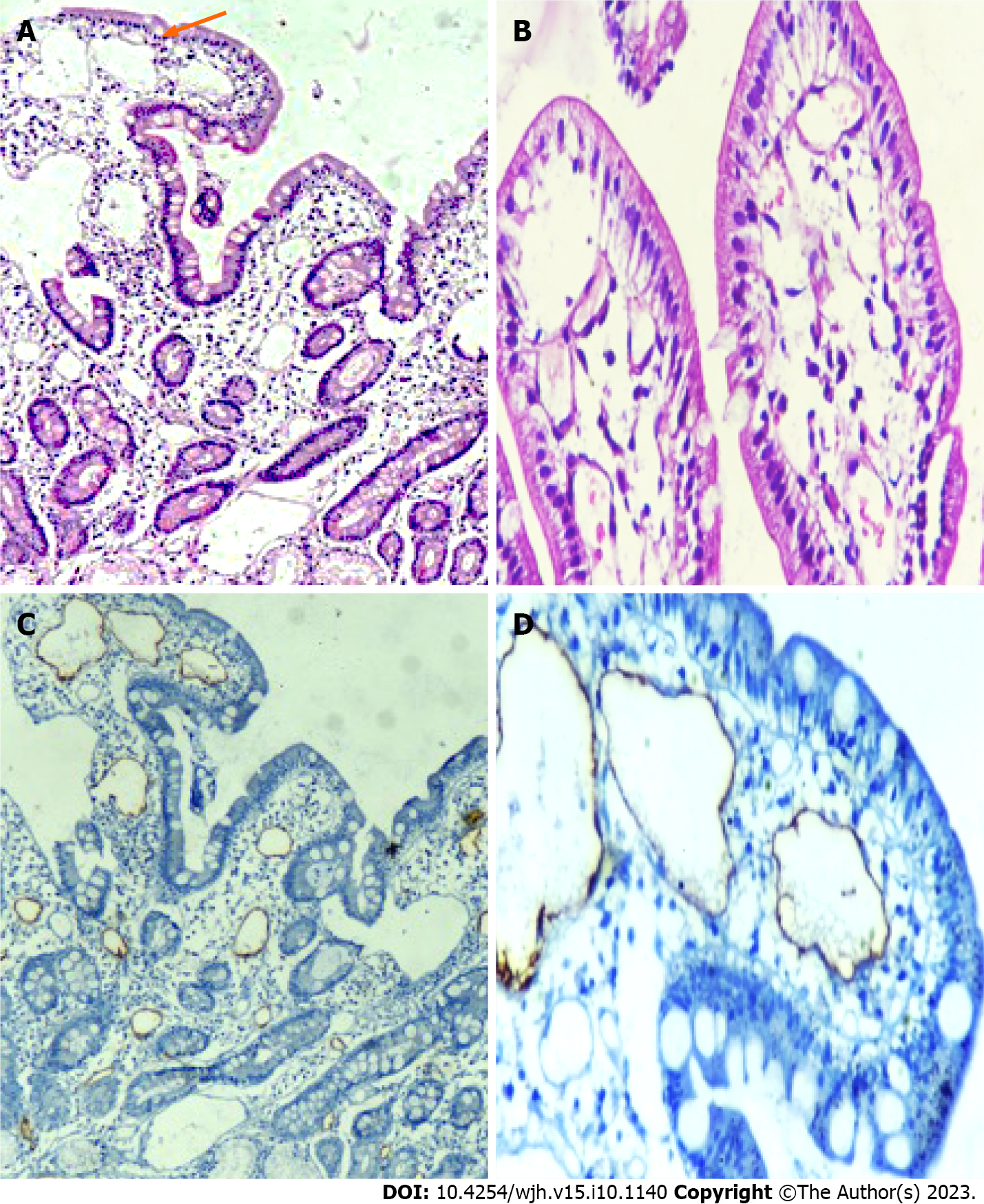
Figure 4 Histological examination of second part of duodenum biopsy specimens.
A-D: Representative histopathological images (hematoxylin and eosin) of duodenum showing markedly dilated vessels in the lamina propria (A and B), which on immunohistochemistry showing strong D2-40 positivity (C and D), confirming the presence of intestinal lymphangiectasia.
- Citation: Arya R, Kumar R, Kumar T, Kumar S, Anand U, Priyadarshi RN, Maji T. Prevalence and risk factors of lymphatic dysfunction in cirrhosis patients with refractory ascites: An often unconsidered mechanism. World J Hepatol 2023; 15(10): 1140-1152
- URL: https://www.wjgnet.com/1948-5182/full/v15/i10/1140.htm
- DOI: https://dx.doi.org/10.4254/wjh.v15.i10.1140


