Copyright
©The Author(s) 2022.
World J Hepatol. Sep 27, 2022; 14(9): 1830-1839
Published online Sep 27, 2022. doi: 10.4254/wjh.v14.i9.1830
Published online Sep 27, 2022. doi: 10.4254/wjh.v14.i9.1830
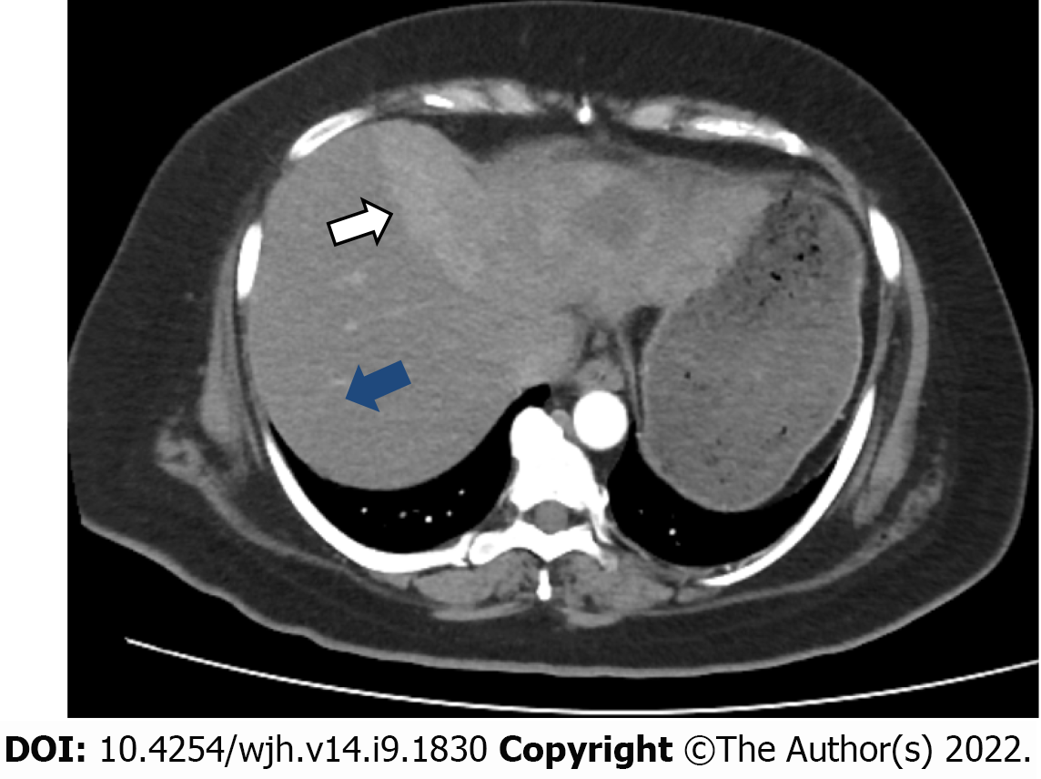
Figure 1 Contrast enhanced tomography scan image demonstrating a large enhancing heterogeneous mass in the left lobe of the liver (white arrow), surrounding normal the liver tissue (blue arrow).
- Citation: Ahmed H, Bari H, Nisar Sheikh U, Basheer MI. Primary hepatic leiomyosarcoma: A case report and literature review . World J Hepatol 2022; 14(9): 1830-1839
- URL: https://www.wjgnet.com/1948-5182/full/v14/i9/1830.htm
- DOI: https://dx.doi.org/10.4254/wjh.v14.i9.1830


