Copyright
©The Author(s) 2022.
World J Hepatol. Sep 27, 2022; 14(9): 1790-1803
Published online Sep 27, 2022. doi: 10.4254/wjh.v14.i9.1790
Published online Sep 27, 2022. doi: 10.4254/wjh.v14.i9.1790
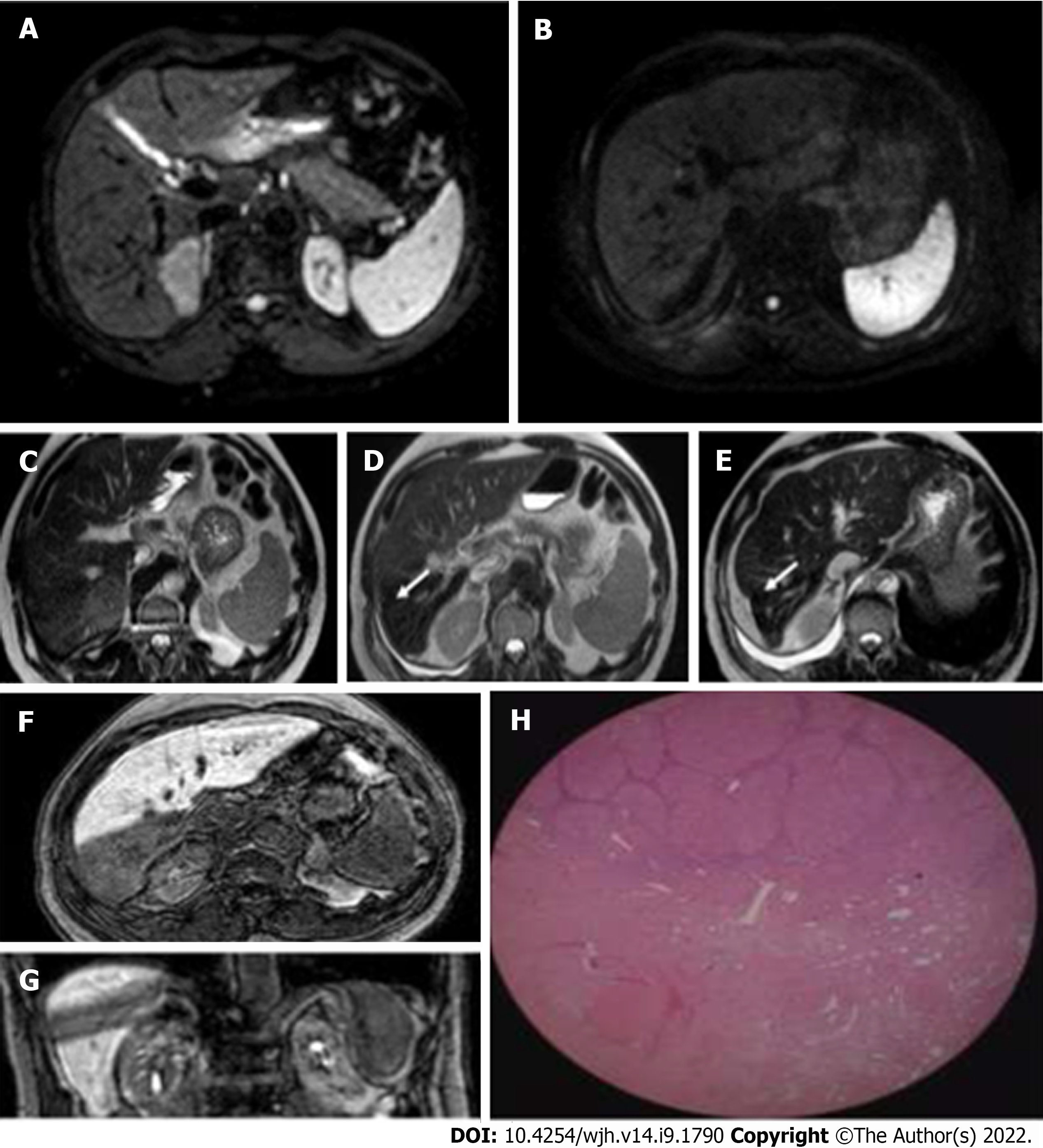
Figure 13 Hepatocellular carcinoma in a 75-year-old man treated with stereotactic body radiation therapy.
A and B: Diffusion-weighted imaging signal of a lesion located in segment VII, before and after treatment. In the pre-therapy evaluation, the nodule presented a marked narrowing of the diffusion of water molecules, a feature no longer present at post-stereotactic body radiation therapy (SBRT) examination; C: Hepatocellular carcinoma nodule located in segment VII characterized by intermediate signal on T2-weighted imaging; D and E: Initial capsular retraction (white arrow) was already evident at the first post-SBRT magnetic resonance examination and particularly evident at the 9 mo follow-up; F and G: SBRT outcomes characterised by an area of "band" hypodensity in the hepatic excretion phase in the axial and coronal sequences, corresponding to shaded hyperintensity in the T2-weighted sequences with the same distribution and morphology; H: These findings are an expression of treatment-induced fibrosis as demonstrated by the histological finding, where postradiotherapy fibrotic tissue can be identified after liver transplantation.
- Citation: Serafini A, Ruggeri V, Inchingolo R, Gatti M, Guarneri A, Maino C, Ippolito D, Grazioli L, Ricardi U, Faletti R. Liver magnetic resonance imaging for evaluation of response to treatment after stereotactic body radiation therapy of hepatocellular carcinoma. World J Hepatol 2022; 14(9): 1790-1803
- URL: https://www.wjgnet.com/1948-5182/full/v14/i9/1790.htm
- DOI: https://dx.doi.org/10.4254/wjh.v14.i9.1790


