Copyright
©The Author(s) 2022.
World J Hepatol. Jul 27, 2022; 14(7): 1520-1527
Published online Jul 27, 2022. doi: 10.4254/wjh.v14.i7.1520
Published online Jul 27, 2022. doi: 10.4254/wjh.v14.i7.1520
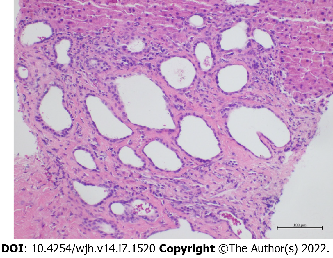
Figure 3 Histopathological examination of the punctured liver biopsy specimen.
Microphotograph of histological appearance of liver biopsy specimen showing in the peri-portal region a group of ductal structures embedded in a hyalinized stroma. The ductal structures appear variably dilated and focal microcystic. These ducts are lined with a cubic or flattened biliary epithelium. (hematoxylin-eosin staining, magnification × 20).
- Citation: Priadko K, Niosi M, Vitale LM, De Sio C, Romano M, De Sio I. “Starry liver” - Von Meyenburg complex clinical case presentation and differential diagnosis discussion: A case report. World J Hepatol 2022; 14(7): 1520-1527
- URL: https://www.wjgnet.com/1948-5182/full/v14/i7/1520.htm
- DOI: https://dx.doi.org/10.4254/wjh.v14.i7.1520


