Copyright
©The Author(s) 2022.
World J Hepatol. Jul 27, 2022; 14(7): 1504-1511
Published online Jul 27, 2022. doi: 10.4254/wjh.v14.i7.1504
Published online Jul 27, 2022. doi: 10.4254/wjh.v14.i7.1504
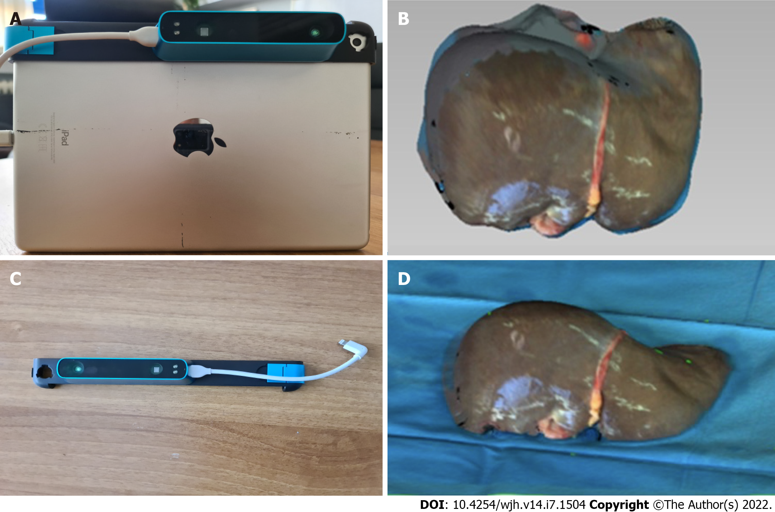
Figure 2 The particular sensor can be adapted to any device with iOS and iPadOS operating system.
A: The device used in the present study. The structure core sensor, the adjustment bracket, the USB communication cable, and the iPad (6th generation) are shown; B: The Occipital original structure sensor; C: An exported 3D model of a liver graft; D: The final 3D model of the liver graft.
- Citation: Katsanos G, Karakasi KE, Karolos IA, Kofinas A, Antoniadis N, Tsioukas V, Tsoulfas G. Volumetric assessment of hepatic grafts using a light detection and ranging system for 3D scanning: Preliminary data. World J Hepatol 2022; 14(7): 1504-1511
- URL: https://www.wjgnet.com/1948-5182/full/v14/i7/1504.htm
- DOI: https://dx.doi.org/10.4254/wjh.v14.i7.1504


