Copyright
©The Author(s) 2022.
World J Hepatol. Jul 27, 2022; 14(7): 1365-1381
Published online Jul 27, 2022. doi: 10.4254/wjh.v14.i7.1365
Published online Jul 27, 2022. doi: 10.4254/wjh.v14.i7.1365
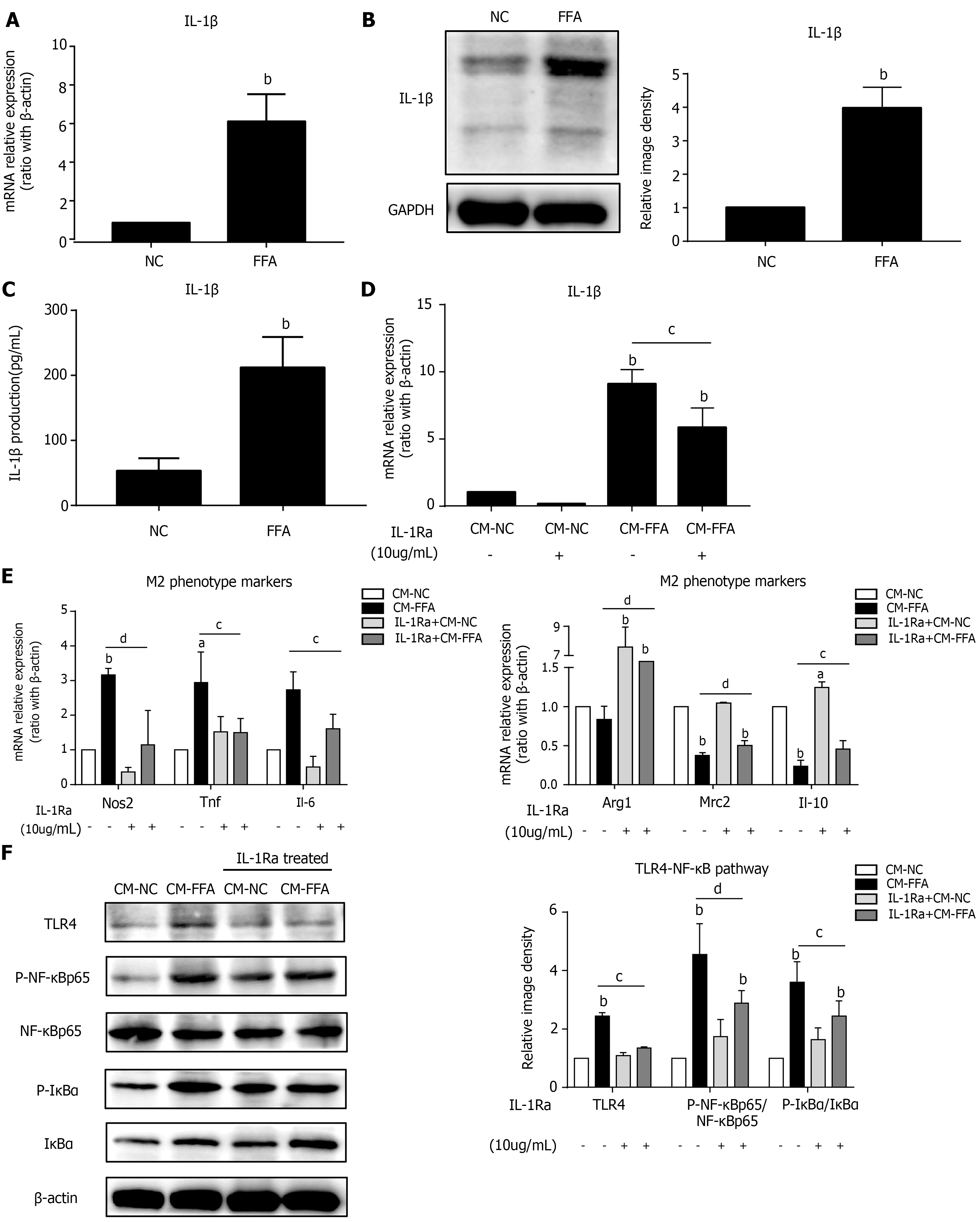
Figure 2 Lipid-laden hepatocytes induce macrophage M1 polarization and inflammation via IL-1β signaling.
Primary hepatocytes were incubated with free fatty acids for 6 h for RT–PCR or 24 h for western blot and ELISA. Cell culture supernatants of hepatocytes were collected and prepared for different conditioned mediums (CMs). RAW264.7 macrophages were pretreated with interleukin-1 receptor antagonist and treated with different CMs for 6 h for RT–PCR or 24 h for western blotting. A: mRNA expression of Il-1β in primary hepatocytes; B: Protein expression of IL-1β in primary hepatocytes; C: IL-1β production in the hepatocyte culture supernatant; D: mRNA expression of Il-1β in macrophages; E: mRNA expression of M1/M2 markers in macrophages; F: Protein expression of the TLR4/NF-κB pathway in macrophages. Values are expressed as the mean ± SE of the mean, aP < 0.05, bP < 0.01 vs normal control (NC) or CM-NC; cP < 0.05, dP < 0.01 comparison of the designated two groups, n = 3 experiments. NC: Normal control; CM: Conditioned medium; GAPDH: Glyceraldehyde-3-phosphate dehydrogenase; IL-1Ra: Interleukin-1 receptor antagonist; Nos: Nitric oxide synthase; Tnf: Tumor necrosis factor; Il: Interleukin; Arg1: Arginine-1; Mrc2: Mannose receptor 2; TLR4: Toll-like receptor 4; NF-κB: Nuclear factor kappa-B; IκBα: Inhibitor of nuclear factor kappa-B.
- Citation: Li XY, Ji PX, Ni XX, Chen YX, Sheng L, Lian M, Guo CJ, Hua J. Regulation of PPAR-γ activity in lipid-laden hepatocytes affects macrophage polarization and inflammation in nonalcoholic fatty liver disease. World J Hepatol 2022; 14(7): 1365-1381
- URL: https://www.wjgnet.com/1948-5182/full/v14/i7/1365.htm
- DOI: https://dx.doi.org/10.4254/wjh.v14.i7.1365


