Copyright
©The Author(s) 2022.
World J Hepatol. Jun 27, 2022; 14(6): 1131-1141
Published online Jun 27, 2022. doi: 10.4254/wjh.v14.i6.1131
Published online Jun 27, 2022. doi: 10.4254/wjh.v14.i6.1131
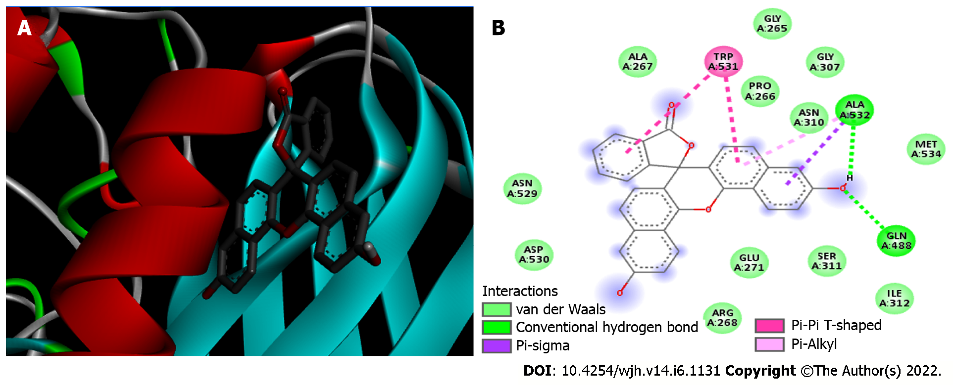
Figure 4 Furin/Naphthofluorescein complex.
A: 3D view of Naphthofluorescein inhibitor (sticks) bound to Furin (Protein Data Bank 5JXI). Furin is shown in blue beta sheets, red alpha-helices, grey random coils and green turns; B: 2D diagram showing the Furin residues that bind to Naphthofluorescein. The internal legend indicates the bond type by color. Amino acid residues are shown in 3-letter code; the number indicates the position within the chain, and the capital letters (A) indicate which chain the residue belongs to.
- Citation: Peiter GC, de Souza CBT, de Oliveira LM, Pagliarin LG, dos Anjos VNF, da Silva FAF, de Melo FF, Teixeira KN. COVID-19 liver and gastroenterology findings: An in silico analysis of SARS-CoV-2 interactions with liver molecules. World J Hepatol 2022; 14(6): 1131-1141
- URL: https://www.wjgnet.com/1948-5182/full/v14/i6/1131.htm
- DOI: https://dx.doi.org/10.4254/wjh.v14.i6.1131


