Copyright
©The Author(s) 2022.
World J Hepatol. Apr 27, 2022; 14(4): 860-865
Published online Apr 27, 2022. doi: 10.4254/wjh.v14.i4.860
Published online Apr 27, 2022. doi: 10.4254/wjh.v14.i4.860
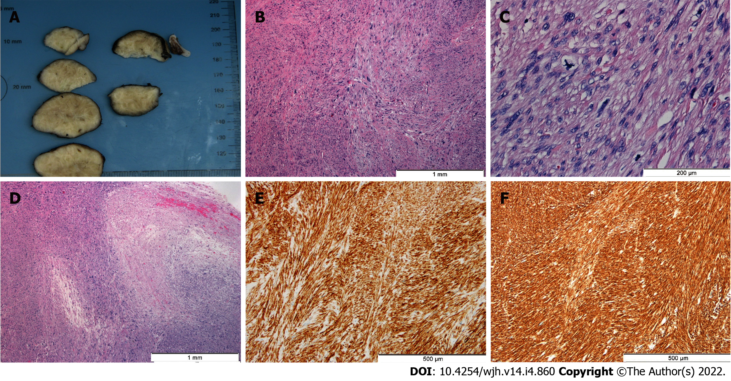
Figure 2 Hepatic leiomyosarcoma.
A: A well-circumscribed, white, solid mass of 4.3 cm exhibiting whorled features was seen on gross examination. B: Histologically, intersecting fascicles of spindle cells with elongate to ovoid nuclei displaying marked pleomorphism were observed [hematoxylin & eosin (HE), 40 ×]. C: Mitotic figures, including atypical ones, were frequent (7 mitosis/10 high power field) (HE, 200 ×). D: Foci of coagulative necrosis were observed (HE, 40 ×). E and F: Tumor cells were diffusely positive for desmin (100 ×) (E) and smooth muscle actin (100 ×) (F), in the absence of S100, c-Kit and discovered on GIST-1 (not shown).
- Citation: Garrido I, Andrade P, Pacheco J, Rios E, Macedo G. Not all liver tumors are alike — an accidentally discovered primary hepatic leiomyosarcoma: A case report. World J Hepatol 2022; 14(4): 860-865
- URL: https://www.wjgnet.com/1948-5182/full/v14/i4/860.htm
- DOI: https://dx.doi.org/10.4254/wjh.v14.i4.860


