Copyright
©The Author(s) 2022.
World J Hepatol. Mar 27, 2022; 14(3): 495-503
Published online Mar 27, 2022. doi: 10.4254/wjh.v14.i3.495
Published online Mar 27, 2022. doi: 10.4254/wjh.v14.i3.495
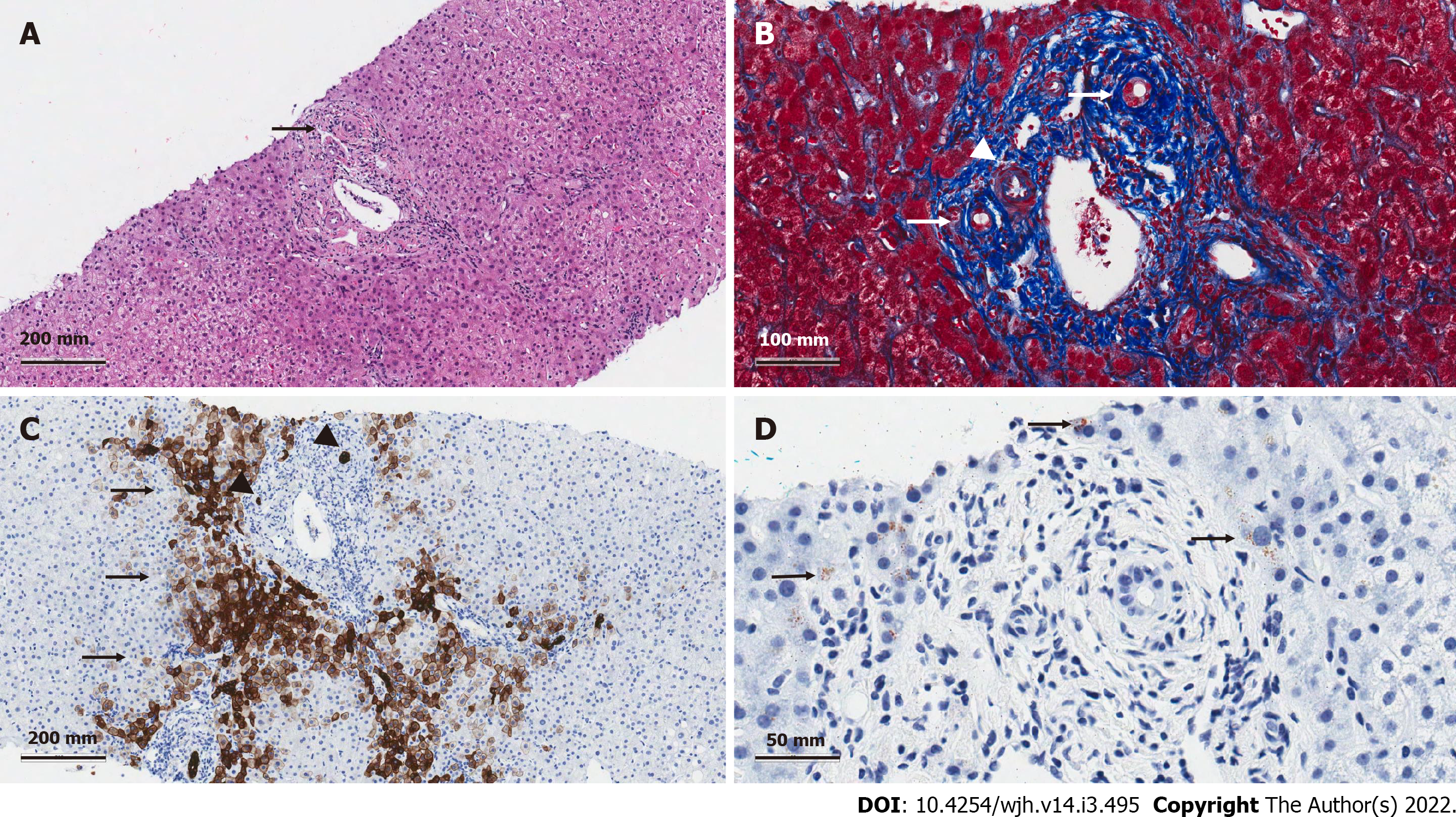
Figure 1 Histologic features in a liver biopsy collected from a patient with normal magnetic resonance cholangiopancreatography and suspected small duct primary sclerosing cholangitis.
A: The H&E shows a portal tract with bile duct senescence (arrow) and mild non-specific, lymphocytic inflammation. B: The Masson’s trichrome stain shows a higher power image of this same portal tract, highlighting the smaller size of the intrahepatic bile ducts when compared to the adjacent hepatic artery (arrowhead). This stain also shows peribiliary sclerosis that is causing fibrous obliteration of the bile duct epithelium (arrows). The collagen surrounding the ducts is dense and has a keloid-like appearance. C: A cytokeratin 7 stain was conducted and shows prominent periportal cholangiolar metaplasia of hepatocytes (arrows) with atrophy of the intrahepatic bile ducts (arrowhead). In patients without chronic biliary obstruction or suboptimal bile flow, cholangiolar metaplasia is not present. D: A copper stain was positive for periportal deposition (arrows), further supporting the presence of chronic biliary obstruction.
- Citation: Nguyen CM, Kline KT, Stevenson HL, Khan K, Parupudi S. Small duct primary sclerosing cholangitis: A discrete variant or a bridge to large duct disease, a practical review. World J Hepatol 2022; 14(3): 495-503
- URL: https://www.wjgnet.com/1948-5182/full/v14/i3/495.htm
- DOI: https://dx.doi.org/10.4254/wjh.v14.i3.495


