Copyright
©The Author(s) 2022.
World J Hepatol. Feb 27, 2022; 14(2): 386-399
Published online Feb 27, 2022. doi: 10.4254/wjh.v14.i2.386
Published online Feb 27, 2022. doi: 10.4254/wjh.v14.i2.386
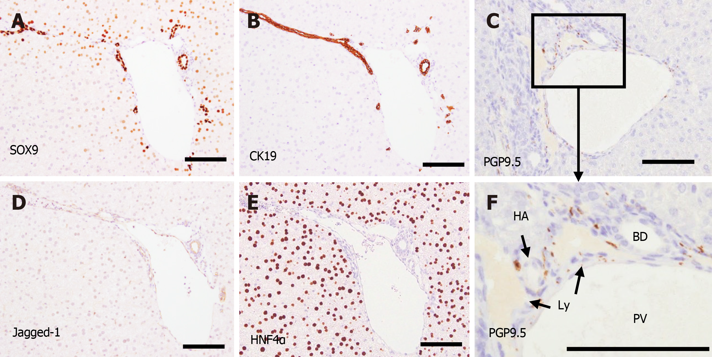
Figure 8 Immunohistochemical analysis of the portal tract in the periphery of the liver at postnatal day 28.
A: SRY-related high mobility group-box gene 9 (SOX9) was expressed in the nuclei of biliary cells; B, C and F: Many cytokeratin 19-positive biliary cells (B) and protein gene product 9.5-positive nerve fibers (C and F) were found in the portal tracts even in the periphery. No nerve fibers were found in the liver parenchyma; D: Jagged-1 expression was mainly found in the bile ducts and vessels; E: Hepatocyte nuclear factor 4α was expressed in the entire liver parenchyma. Scale bars = 200 μm. PV: Portal vein; HA: Hepatic artery; BD: Bile duct; Ly: Lymphatic vessel; CK19: Cytokeratin 19; SOX9: SRY-related high mobility group-box gene 9; PGP9.5: Protein gene product 9.5.
- Citation: Koike N, Tadokoro T, Ueno Y, Okamoto S, Kobayashi T, Murata S, Taniguchi H. Development of the nervous system in mouse liver. World J Hepatol 2022; 14(2): 386-399
- URL: https://www.wjgnet.com/1948-5182/full/v14/i2/386.htm
- DOI: https://dx.doi.org/10.4254/wjh.v14.i2.386


