Copyright
©The Author(s) 2022.
World J Hepatol. Feb 27, 2022; 14(2): 386-399
Published online Feb 27, 2022. doi: 10.4254/wjh.v14.i2.386
Published online Feb 27, 2022. doi: 10.4254/wjh.v14.i2.386
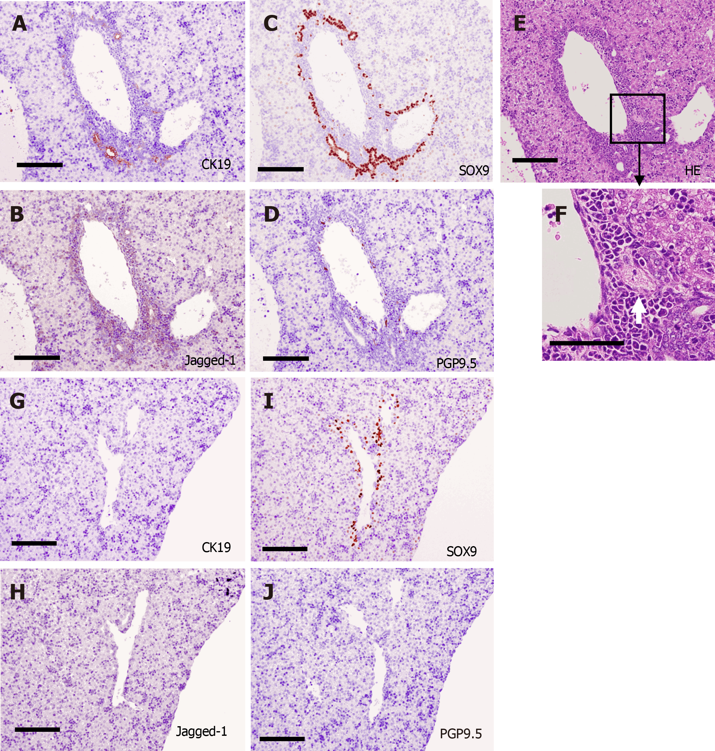
Figure 6 Immunohistochemical analysis of the area around the portal vein at postnatal day 0.
A-F: Center of the liver; A-C: Many SRY-related high mobility group-box gene 9 (SOX9)-positive cells were found in Jagged-1-positive periportal tissue, and they formed cytokeratin 19 (CK19)-positive bile ducts in the center; D: Protein gene product 9.5 (PGP9.5)-positive nerve fibers were found in the periportal tissue in the center; E and F: Branching of the hepatic artery was found in the periportal tissue (white arrow); G-J: Periphery of the liver. The Jagged-1-positive periportal tissue was thinner in the periphery than in the center, and the number of biliary structures was smaller in the periphery. Neither CK19-positive bile ducts nor PGP9.5-positive nerve fibers were found in the periphery. Scale bars = 200 μm. HE: Hematoxylin-eosin; CK19: Cytokeratin 19; SOX9: SRY-related high mobility group-box gene 9; PGP9.5: Protein gene product 9.5.
- Citation: Koike N, Tadokoro T, Ueno Y, Okamoto S, Kobayashi T, Murata S, Taniguchi H. Development of the nervous system in mouse liver. World J Hepatol 2022; 14(2): 386-399
- URL: https://www.wjgnet.com/1948-5182/full/v14/i2/386.htm
- DOI: https://dx.doi.org/10.4254/wjh.v14.i2.386


