Copyright
©The Author(s) 2022.
World J Hepatol. Feb 27, 2022; 14(2): 386-399
Published online Feb 27, 2022. doi: 10.4254/wjh.v14.i2.386
Published online Feb 27, 2022. doi: 10.4254/wjh.v14.i2.386
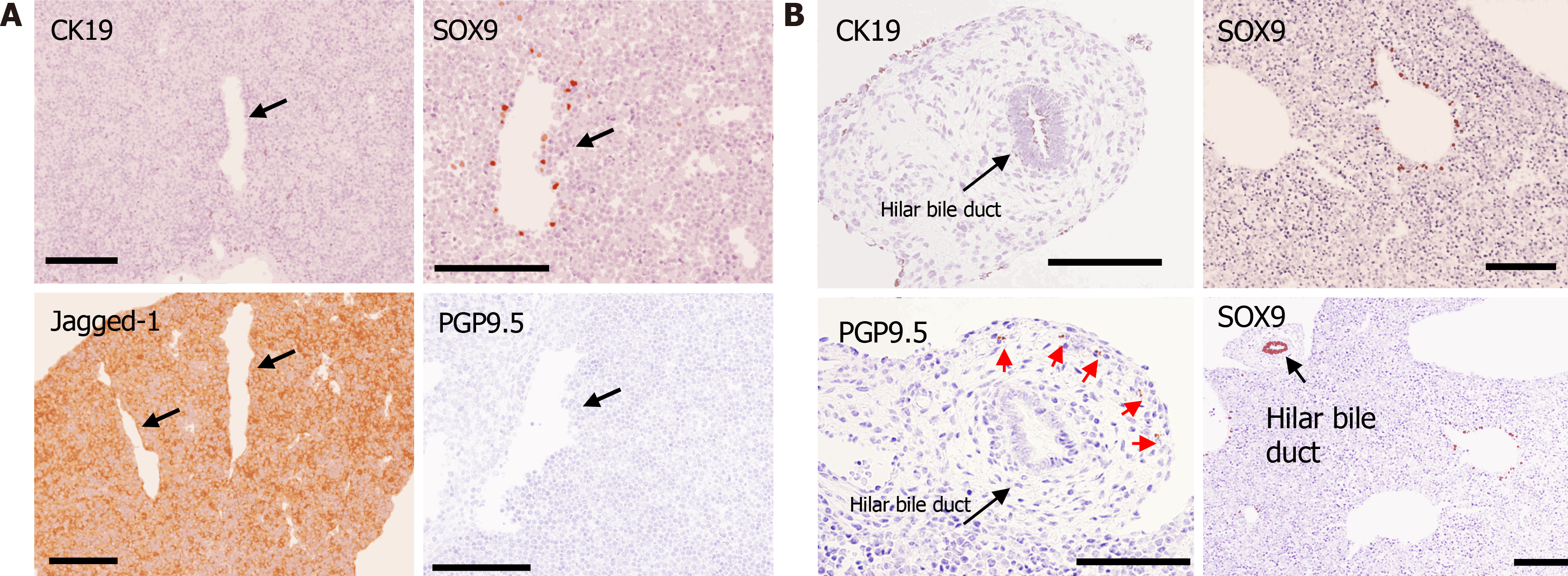
Figure 4 Immunohistochemical analysis.
A: Immunohistochemical analysis of the liver at embryonic day 12.5. Jagged-1 was diffusely positive in the liver (lower left panel), and SRY-related high mobility group-box gene 9 (SOX9)-positive bile duct progenitor cells were found around the large vessels presumably defined as portal veins (upper right panel); however, cytokeratin 19 (CK19) (another marker for bile duct)- and protein gene product 9.5 (PGP9.5) (a marker for neurons)-positive cells were not found (shown in the upper left panel and lower right panels, respectively). Arrows indicate portal veins; B: Immunohistochemical analysis of the center of the liver at embryonic day 15.5. SOX9-positive bile duct progenitor cells were found around the large vessel presumably defined as portal veins. The hilar bile duct strongly expressed SOX9 and weakly expressed CK19. PGP9.5-positive bodies were only found in the parenchyma around the hilar bile duct (red arrow). Scale bars = 200 μm. CK19: Cytokeratin 19; SOX9: SRY-related high mobility group-box gene 9; PGP9.5: Protein gene product 9.5.
- Citation: Koike N, Tadokoro T, Ueno Y, Okamoto S, Kobayashi T, Murata S, Taniguchi H. Development of the nervous system in mouse liver. World J Hepatol 2022; 14(2): 386-399
- URL: https://www.wjgnet.com/1948-5182/full/v14/i2/386.htm
- DOI: https://dx.doi.org/10.4254/wjh.v14.i2.386


