Copyright
©The Author(s) 2022.
World J Hepatol. Feb 27, 2022; 14(2): 386-399
Published online Feb 27, 2022. doi: 10.4254/wjh.v14.i2.386
Published online Feb 27, 2022. doi: 10.4254/wjh.v14.i2.386
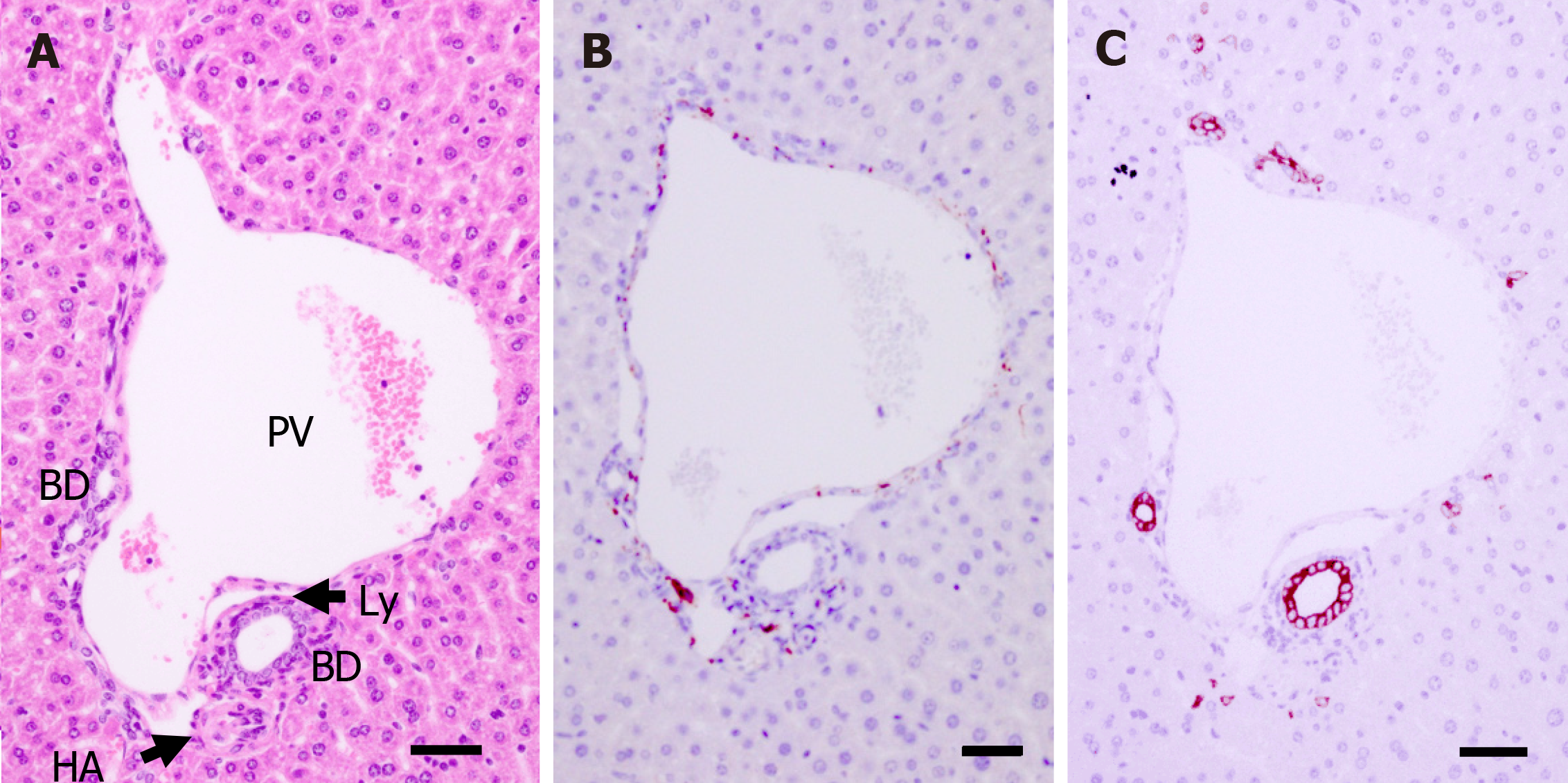
Figure 2 Histology of the portal tract of a postnatal day 56 mouse.
A: Morphological analysis of a portal tract via hematoxylin-eosin staining; B and C: Neurons and bile ducts formed around the portal tract are shown by protein gene product 9.5 staining (B) and cytokeratin 19 staining (C), respectively. PV: Portal vein; HA: Hepatic artery; BD: Bile duct; Ly: Lymphatic vessel. Scale bar = 50 μm.
- Citation: Koike N, Tadokoro T, Ueno Y, Okamoto S, Kobayashi T, Murata S, Taniguchi H. Development of the nervous system in mouse liver. World J Hepatol 2022; 14(2): 386-399
- URL: https://www.wjgnet.com/1948-5182/full/v14/i2/386.htm
- DOI: https://dx.doi.org/10.4254/wjh.v14.i2.386


