Copyright
©The Author(s) 2021.
World J Hepatol. Sep 27, 2021; 13(9): 1079-1097
Published online Sep 27, 2021. doi: 10.4254/wjh.v13.i9.1079
Published online Sep 27, 2021. doi: 10.4254/wjh.v13.i9.1079
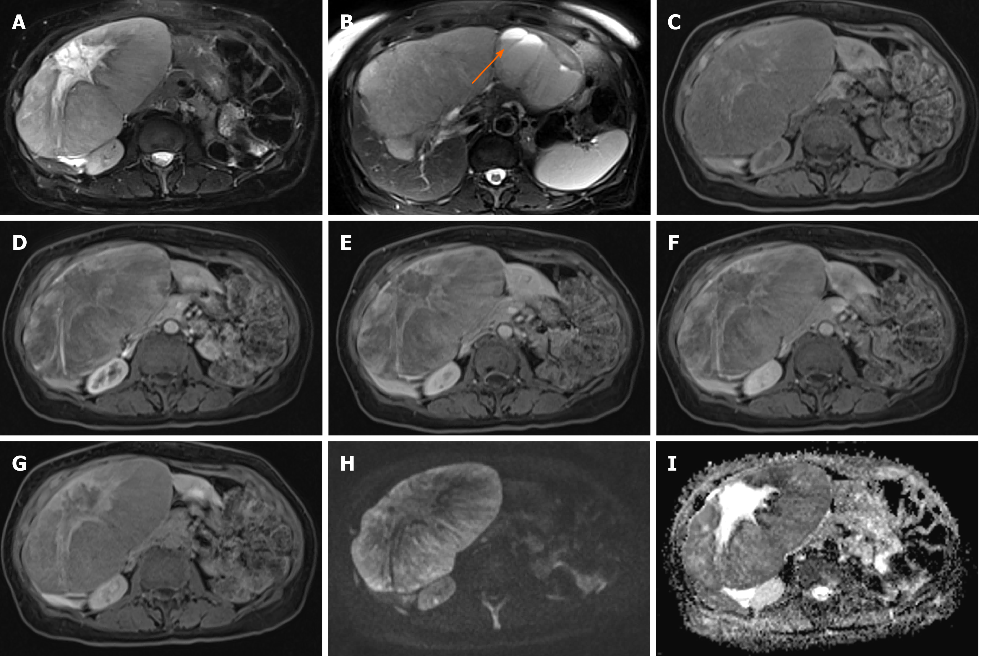
Figure 13 Neuroendocrine carcinoma.
A 69-year-old female was found to have incidental large liver lesions in a non-cirrhotic liver while undergoing magnetic resonance (MR) pelvis for a uterine lesion, presumed to be fibroid. A: MR demonstrated large liver masses, the largest exophytic mass showing intermediate to high T2 signal with a high signal stellate scar; B: One of the lesions in the left liver lobe demonstrates a cystic component with fluid-fluid levels, which was presumed to represent previous haemorrhage; C: Majority of the lesions were of low T1 signal with a few hyperintense flecks surrounding the scar; D-F: There was heterogenous enhancement on arterial phase (D) with no washout demonstrated on portal venous (E) and delayed (F) phases; G: Hepatobiliary phase showed no contrast retention within the lesion except for the central scar; H and I: Diffusion weighted imaging at b800 (H) and apparent diffusion coefficient (I) show areas of diffusion restriction. These were biopsied and histology demonstrated well differentiated neuroendocrine carcinoma. The origin of this was not determinable from the immunohistochemical pattern. Overall, this was favoured to represent a primary neuroendocrine tumour of the liver as further imaging did not reveal another primary (although admittedly biopsy of the uterine lesion, radiologically presumed fibroid, was never performed). The patient represented a month later with haemorrhagic brain metastases.
- Citation: Noreikaite J, Albasha D, Chidambaram V, Arora A, Katti A. Indeterminate liver lesions on gadoxetic acid-enhanced magnetic resonance imaging of the liver: Case-based radiologic-pathologic review. World J Hepatol 2021; 13(9): 1079-1097
- URL: https://www.wjgnet.com/1948-5182/full/v13/i9/1079.htm
- DOI: https://dx.doi.org/10.4254/wjh.v13.i9.1079


