Copyright
©The Author(s) 2021.
World J Hepatol. Sep 27, 2021; 13(9): 1079-1097
Published online Sep 27, 2021. doi: 10.4254/wjh.v13.i9.1079
Published online Sep 27, 2021. doi: 10.4254/wjh.v13.i9.1079
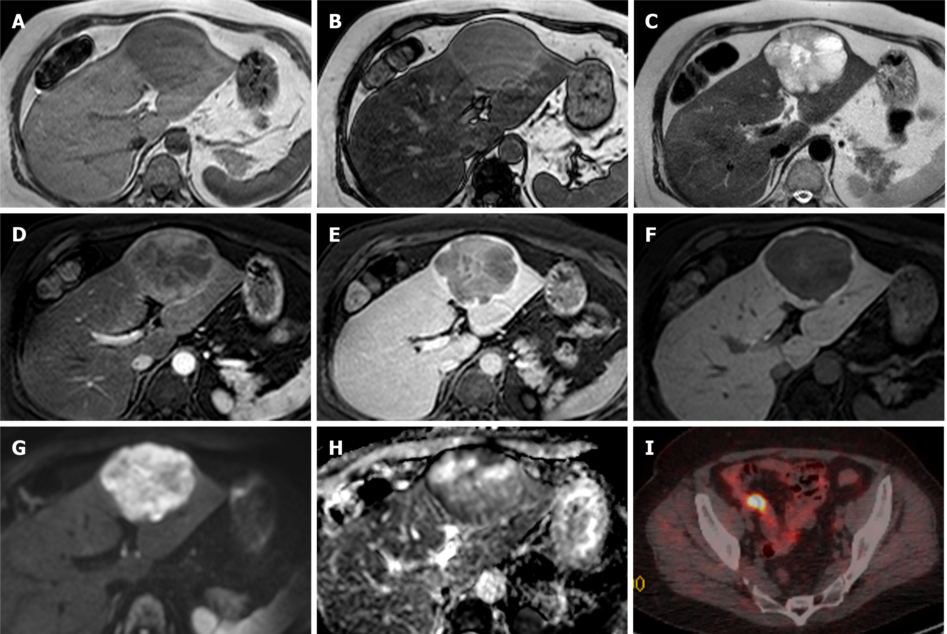
Figure 12 Neuroendocrine carcinoma metastases.
A 59-year-old female was found to have a few liver lesions, the dominant lesion in the left lobe demonstrated here. A and B: In-phase (A) and out-of-phase (B) sequences show background hepatic steatosis, but no tumoral fat; C: The lesion shows heterogenous high T2 signal; D and E: There is mainly peripheral enhancement on the arterial phase (D) with washout on delayed phase (E). Delayed phase also shows an enhancing capsule; F: On hepatobiliary phase there is no contrast retention within the lesion except for the thin-rim of presumed capsule; G and H: There is high signal on diffusion weighted imaging b500 (G) with low signal seen on apparent diffusion coefficient (H), especially in the periphery. The other smaller lesions (not demonstrated here) showed similar signal characteristics. Initial staging computed tomography showed no primary tumour to suggest this is metastasis. The lesions were resected and histology confirmed low grade neuroendocrine tumour, with Ki-67 proliferation index of less than 1%; I: The patient underwent subsequent positron emission tomography scan that demonstrated the primary in the distal ileum.
- Citation: Noreikaite J, Albasha D, Chidambaram V, Arora A, Katti A. Indeterminate liver lesions on gadoxetic acid-enhanced magnetic resonance imaging of the liver: Case-based radiologic-pathologic review. World J Hepatol 2021; 13(9): 1079-1097
- URL: https://www.wjgnet.com/1948-5182/full/v13/i9/1079.htm
- DOI: https://dx.doi.org/10.4254/wjh.v13.i9.1079


