Copyright
©The Author(s) 2021.
World J Hepatol. Sep 27, 2021; 13(9): 1079-1097
Published online Sep 27, 2021. doi: 10.4254/wjh.v13.i9.1079
Published online Sep 27, 2021. doi: 10.4254/wjh.v13.i9.1079
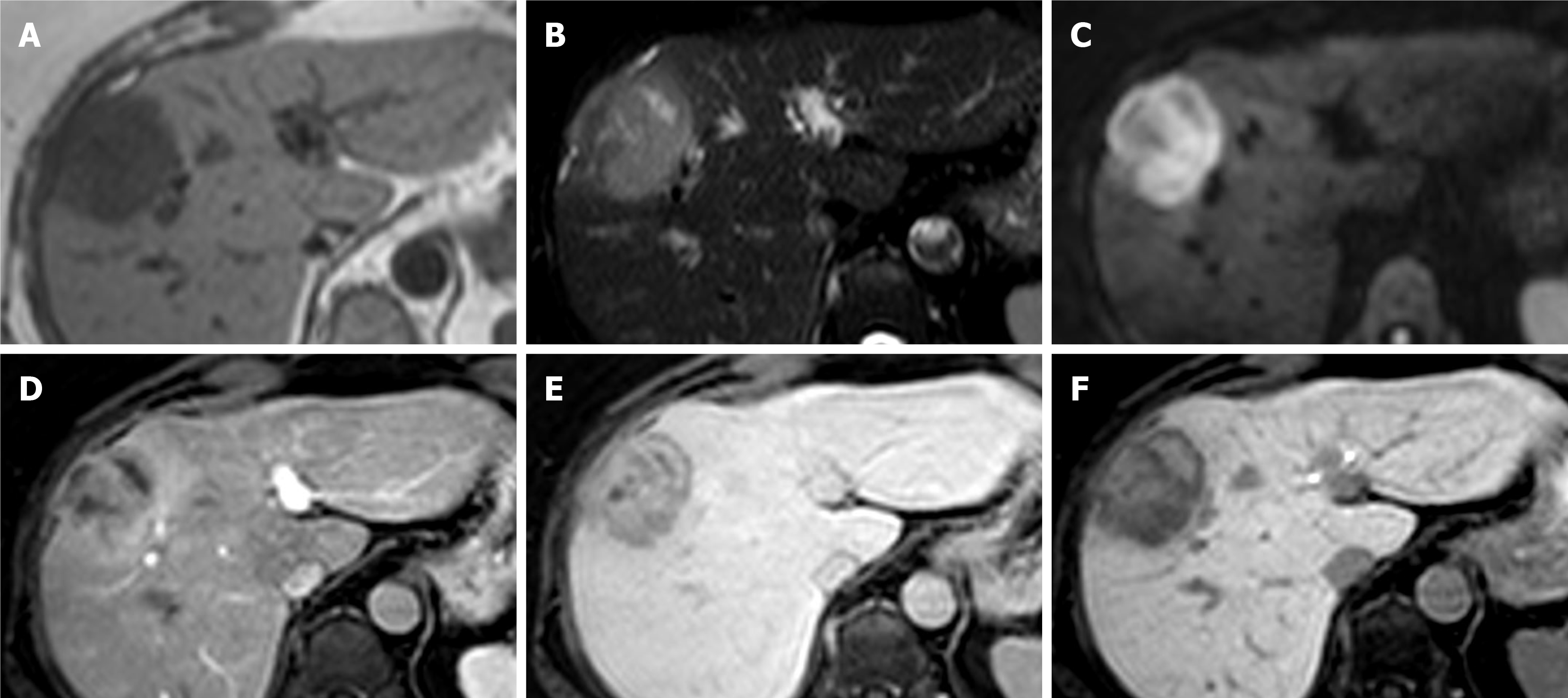
Figure 10 Intrahepatic cholangiocarcinoma.
A 64-year-old female with background of hepatitis C cirrhosis was found to have a liver lesion on surveillance ultrasound. Initial magnetic resonance (MR) with extracellular contrast material was reported as likely hepatocellular carcinoma or metastasis. Biopsy confirmed cholangiocarcinoma and gadoxetic acid enhanced MR was organised to exclude satellite lesions and intrahepatic metastases. A-C: MR shows a right liver lobe lesion which is hypointense on T1-weighted imaging (A), hyperintense on T2-weighted imaging (B) and shows diffusion restriction on b800 diffusion-weighted imaging (C); D and E: On arterial phase (D) there is peripheral enhancement with progressive centripetal enhancement on delayed phases (E); F: Hepatobiliary phase shows a hypointense rim with a cloud-like inhomogeneous central enhancement. No further malignant liver lesions demonstrated.
- Citation: Noreikaite J, Albasha D, Chidambaram V, Arora A, Katti A. Indeterminate liver lesions on gadoxetic acid-enhanced magnetic resonance imaging of the liver: Case-based radiologic-pathologic review. World J Hepatol 2021; 13(9): 1079-1097
- URL: https://www.wjgnet.com/1948-5182/full/v13/i9/1079.htm
- DOI: https://dx.doi.org/10.4254/wjh.v13.i9.1079


