Copyright
©The Author(s) 2021.
World J Hepatol. Sep 27, 2021; 13(9): 1079-1097
Published online Sep 27, 2021. doi: 10.4254/wjh.v13.i9.1079
Published online Sep 27, 2021. doi: 10.4254/wjh.v13.i9.1079
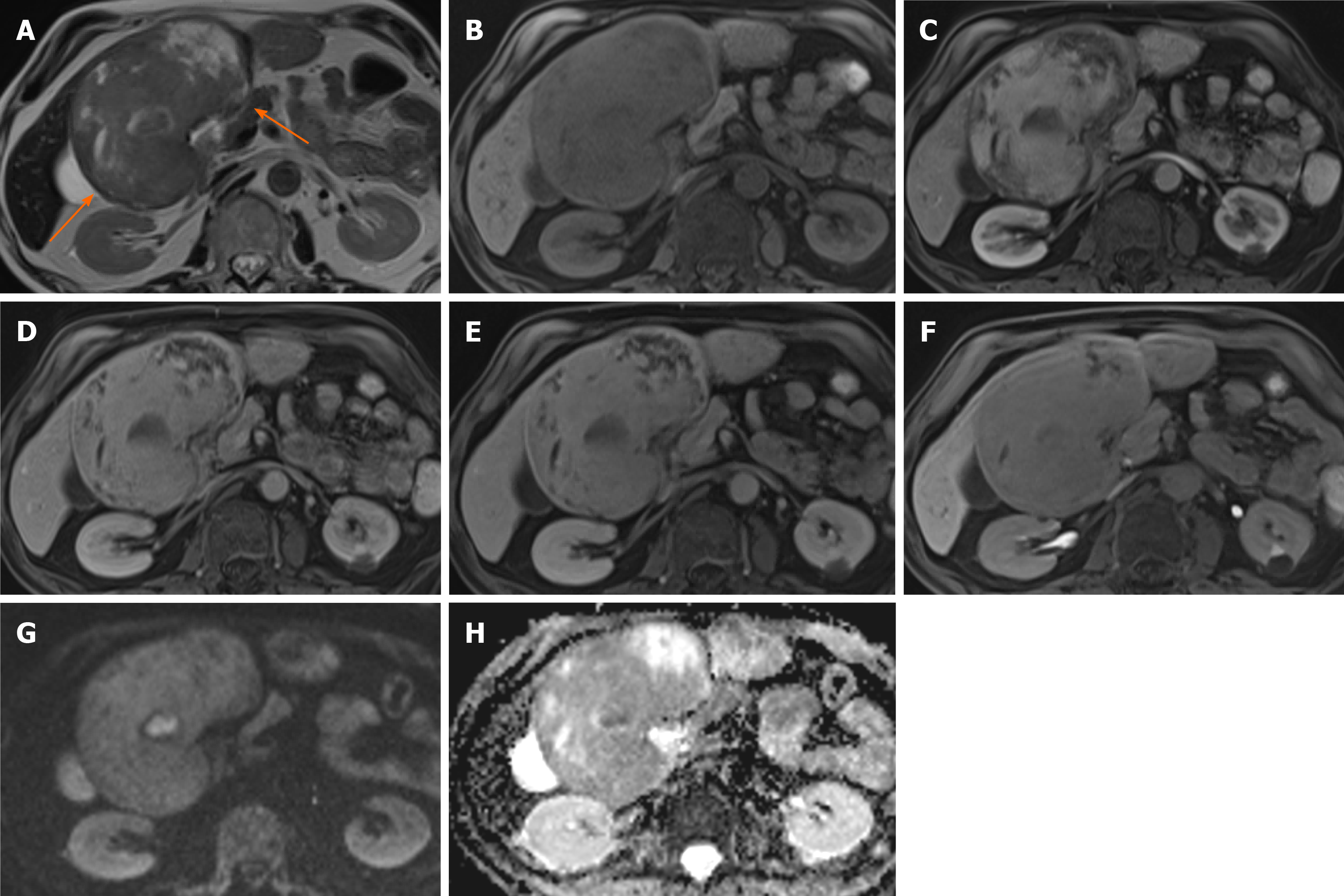
Figure 9 Hepatocellular carcinoma.
A 70-year-old man with a transient episode of frank haematuria as part of the investigations into this, was incidentally found to have a large liver mass arising from the left lobe of the liver. He had previous history of tongue cancer. Liver function tests were normal and alpha-fetoprotein was 2 throughout. A: The lesion (arrowed) is mostly hypointense on T2-weighted sequence with heterogenous areas of high signal; B and C: On T1-weighted sequence (B) it shows iso- to hypointense signal and there is heterogenous arterial enhancement (C); D and E: There is some further filling in on portal venous phase (D) where the lesion is now isointense to the liver parenchyma, similarly to delayed phase (E); F: On hepatobiliary phase the mass is hypointense to background liver; G and H: Diffusion-weighted imaging sequence (G) at b value of 800 shows a focal nodule within the lesion that is markedly hyperintense and on apparent diffusion coefficient (H) hypointense in keeping with diffusion restriction. The lesion was resected and histology confirmed moderately differentiated hepatocellular carcinoma.
- Citation: Noreikaite J, Albasha D, Chidambaram V, Arora A, Katti A. Indeterminate liver lesions on gadoxetic acid-enhanced magnetic resonance imaging of the liver: Case-based radiologic-pathologic review. World J Hepatol 2021; 13(9): 1079-1097
- URL: https://www.wjgnet.com/1948-5182/full/v13/i9/1079.htm
- DOI: https://dx.doi.org/10.4254/wjh.v13.i9.1079


