Copyright
©The Author(s) 2021.
World J Hepatol. Sep 27, 2021; 13(9): 1079-1097
Published online Sep 27, 2021. doi: 10.4254/wjh.v13.i9.1079
Published online Sep 27, 2021. doi: 10.4254/wjh.v13.i9.1079
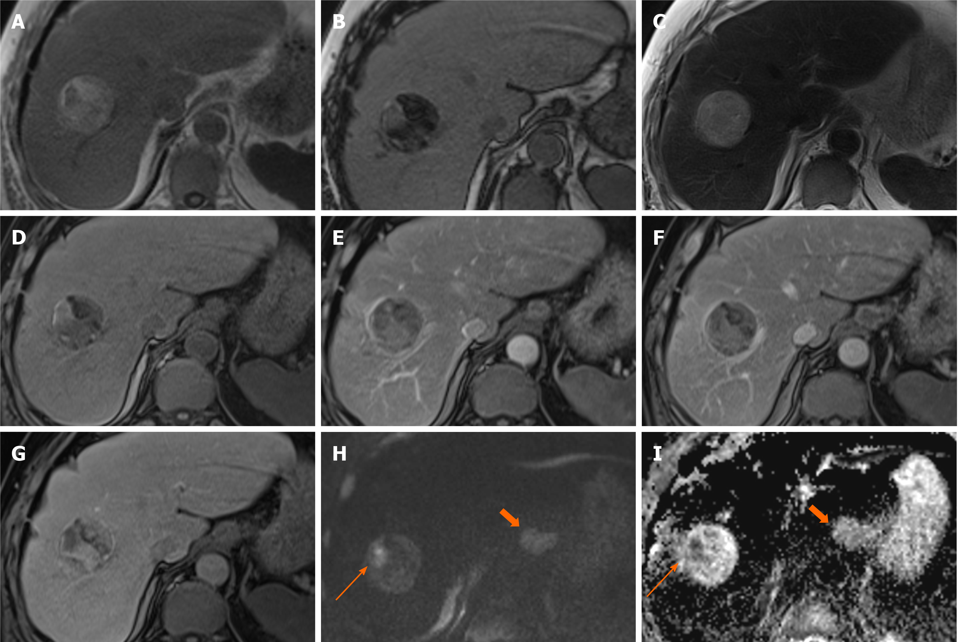
Figure 8 Hepatocellular carcinoma.
A 71-year-old underwent computed tomography chest, abdomen and pelvis for anaemia which identified ascending colon thickening and a liver lesion. Colonoscopy confirmed malignant lesion in the ascending colon and histology showed this to be an adenocarcinoma. Magnetic resonance of liver was performed to characterise the liver mass. A and B: This demonstrates a well-defined lesion with the majority of it showing fat component [signal loss on out-of-phase (B) compared to in-phase (A)] except for a small part laterally; C: It is of mildly high signal on T2 sequences; D: Unenhanced sequence; E-G: There are areas of patchy enhancement on arterial (E) and portal venous (F) phases with heterogenous contrast retention on hepatobiliary phase (G); H and I: This part also shows marked diffusion restriction (long arrow, H–diffusion-weighted imaging b800, I–apparent diffusion coefficient). Diffusion sequences also identified a lymph node showing restricted diffusion (short arrow). Subsequent endoscopy was organised which demonstrated an oesophageal lesion, and biopsies of this, and the adjacent lymph node proved it to be a squamous cell carcinoma. Even with two other primaries, the liver lesion was not considered typical for a metastasis radiologically and targeted biopsy was performed. Histology showed well to moderately differentiated hepatocellular carcinoma.
- Citation: Noreikaite J, Albasha D, Chidambaram V, Arora A, Katti A. Indeterminate liver lesions on gadoxetic acid-enhanced magnetic resonance imaging of the liver: Case-based radiologic-pathologic review. World J Hepatol 2021; 13(9): 1079-1097
- URL: https://www.wjgnet.com/1948-5182/full/v13/i9/1079.htm
- DOI: https://dx.doi.org/10.4254/wjh.v13.i9.1079


