Copyright
©The Author(s) 2021.
World J Hepatol. Sep 27, 2021; 13(9): 1079-1097
Published online Sep 27, 2021. doi: 10.4254/wjh.v13.i9.1079
Published online Sep 27, 2021. doi: 10.4254/wjh.v13.i9.1079
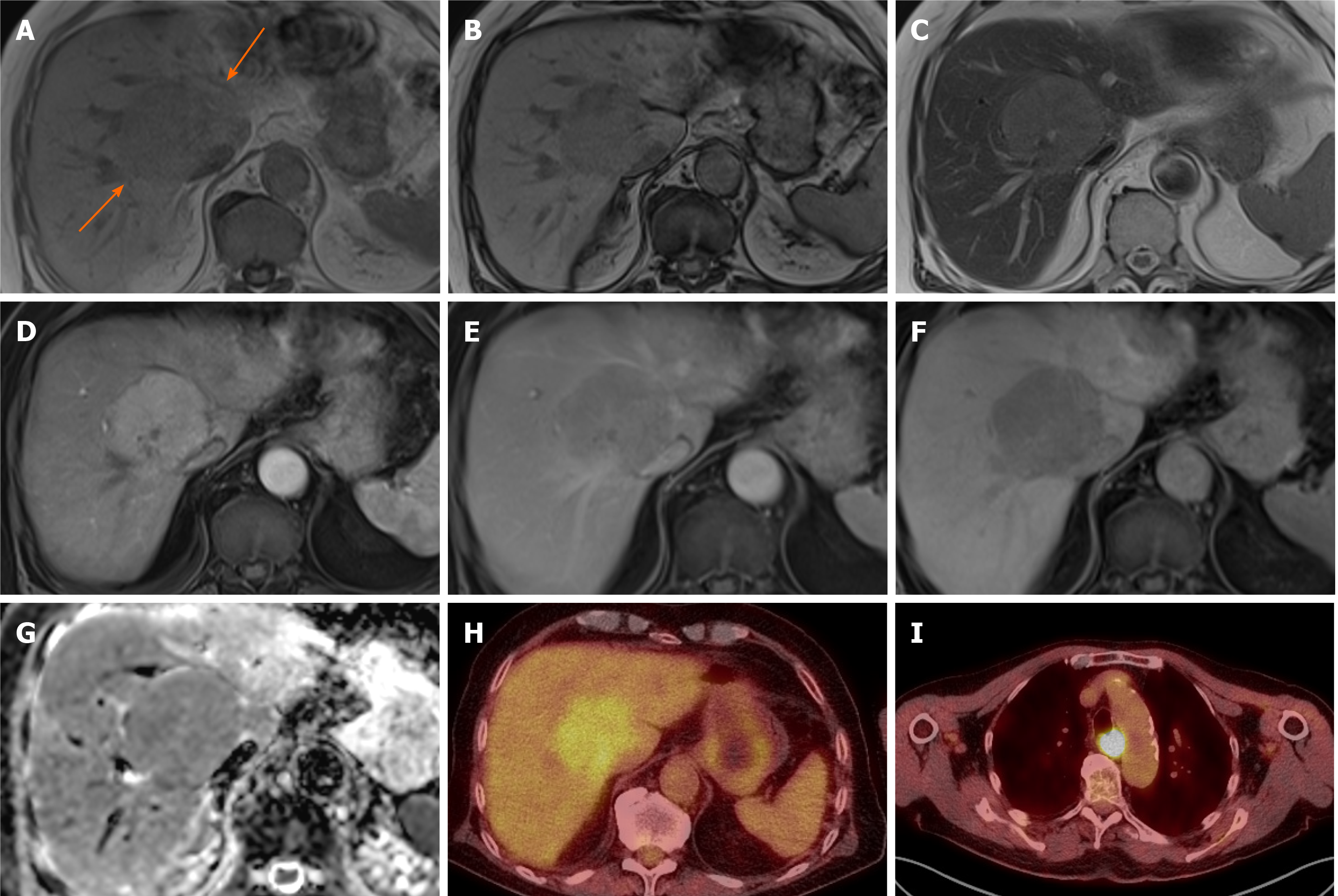
Figure 7 Hepatocellular carcinoma.
A 79-year-old with previous prostate cancer has undergone a magnetic resonance (MR) pelvis and was found to have prostatic cancer recurrence and a liver mass. He has undergone staging computed tomography which showed a further area of oesophageal thickening. Endoscopy revealed oesophageal tumour and biopsy confirmed this to be a squamous cell carcinoma. MR liver and positron emission tomography (PET) scan were performed to characterise these and determine whether liver lesion is a metastasis from oesophageal or prostate primary. Alpha-fetoprotein value was 10 at time of diagnosis. A and B: In- (A) and out-of-phase (B) sequences show low T1 signal liver mass with no intratumoral fat; C: It is of mildly high signal on T2 sequences; D and E: There is homogenous arterial enhancement (D) with washout on portal venous (E) phase; F and G: No contrast retention on hepatobiliary phase (F) and isointense to low signal on apparent diffusion coefficient (G); H and I: PET scan shows tracer uptake within the liver lesion (H), however this is of lower standardized uptake value than the oesophageal cancer (I). Targeted liver lesion biopsy confirmed this to be a hepatocellular carcinoma.
- Citation: Noreikaite J, Albasha D, Chidambaram V, Arora A, Katti A. Indeterminate liver lesions on gadoxetic acid-enhanced magnetic resonance imaging of the liver: Case-based radiologic-pathologic review. World J Hepatol 2021; 13(9): 1079-1097
- URL: https://www.wjgnet.com/1948-5182/full/v13/i9/1079.htm
- DOI: https://dx.doi.org/10.4254/wjh.v13.i9.1079


