Copyright
©The Author(s) 2021.
World J Hepatol. Sep 27, 2021; 13(9): 1079-1097
Published online Sep 27, 2021. doi: 10.4254/wjh.v13.i9.1079
Published online Sep 27, 2021. doi: 10.4254/wjh.v13.i9.1079
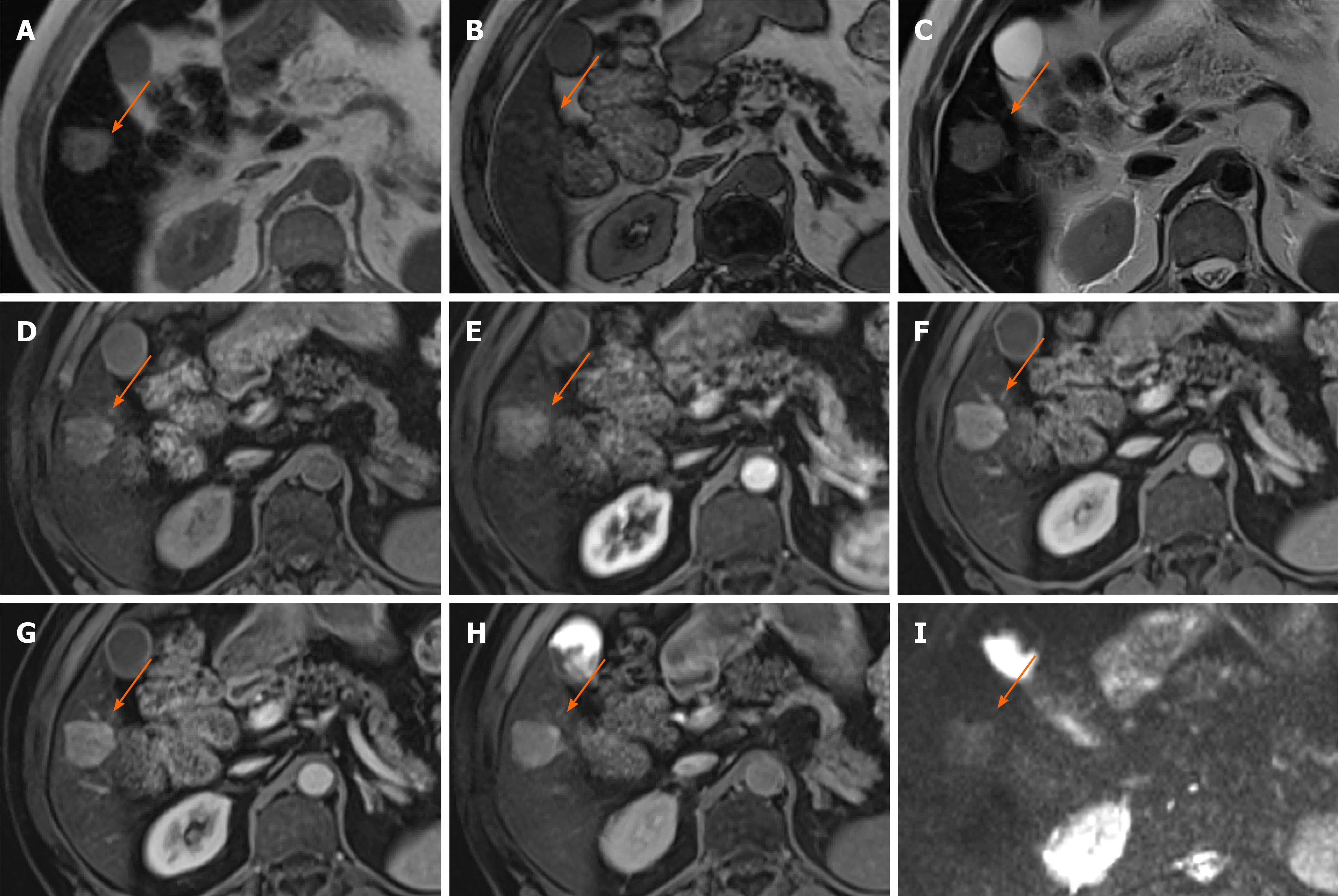
Figure 6 Hepatocellular carcinoma.
A 80-year-old man presented with haematuria and was found to have an incidental liver lesion on computed tomography. His liver function tests were normal. A and B: Magnetic resonance demonstrates signal loss throughout the liver, with paradoxical increase in signal on out-of-phase (B) imaging when compared to in-phase (A), suggestive of underlying iron overload; C: Segment 5 liver lesion shows signal loss on out-of-phase sequences suggesting fat contents and is of high T1 and T2 signal; D: Pre-contrast images; E-G: Subtraction sequences were not performed, but allowing for this, there is some enhancement on arterial phase (E), which persists into portal venous (F) and delayed phases (G); H and I: There is contrast retention on hepatobiliary phase (H) and no diffusion restriction (I–b400). Further tests performed confirmed genetic hemochromatosis. Portal venous pressure measurement also showed portal hypertension. Lesional biopsy confirmed this to be a moderately differentiated hepatocellular carcinoma in a background of cirrhosis, which was subsequently ablated.
- Citation: Noreikaite J, Albasha D, Chidambaram V, Arora A, Katti A. Indeterminate liver lesions on gadoxetic acid-enhanced magnetic resonance imaging of the liver: Case-based radiologic-pathologic review. World J Hepatol 2021; 13(9): 1079-1097
- URL: https://www.wjgnet.com/1948-5182/full/v13/i9/1079.htm
- DOI: https://dx.doi.org/10.4254/wjh.v13.i9.1079


