Copyright
©The Author(s) 2021.
World J Hepatol. Sep 27, 2021; 13(9): 1079-1097
Published online Sep 27, 2021. doi: 10.4254/wjh.v13.i9.1079
Published online Sep 27, 2021. doi: 10.4254/wjh.v13.i9.1079
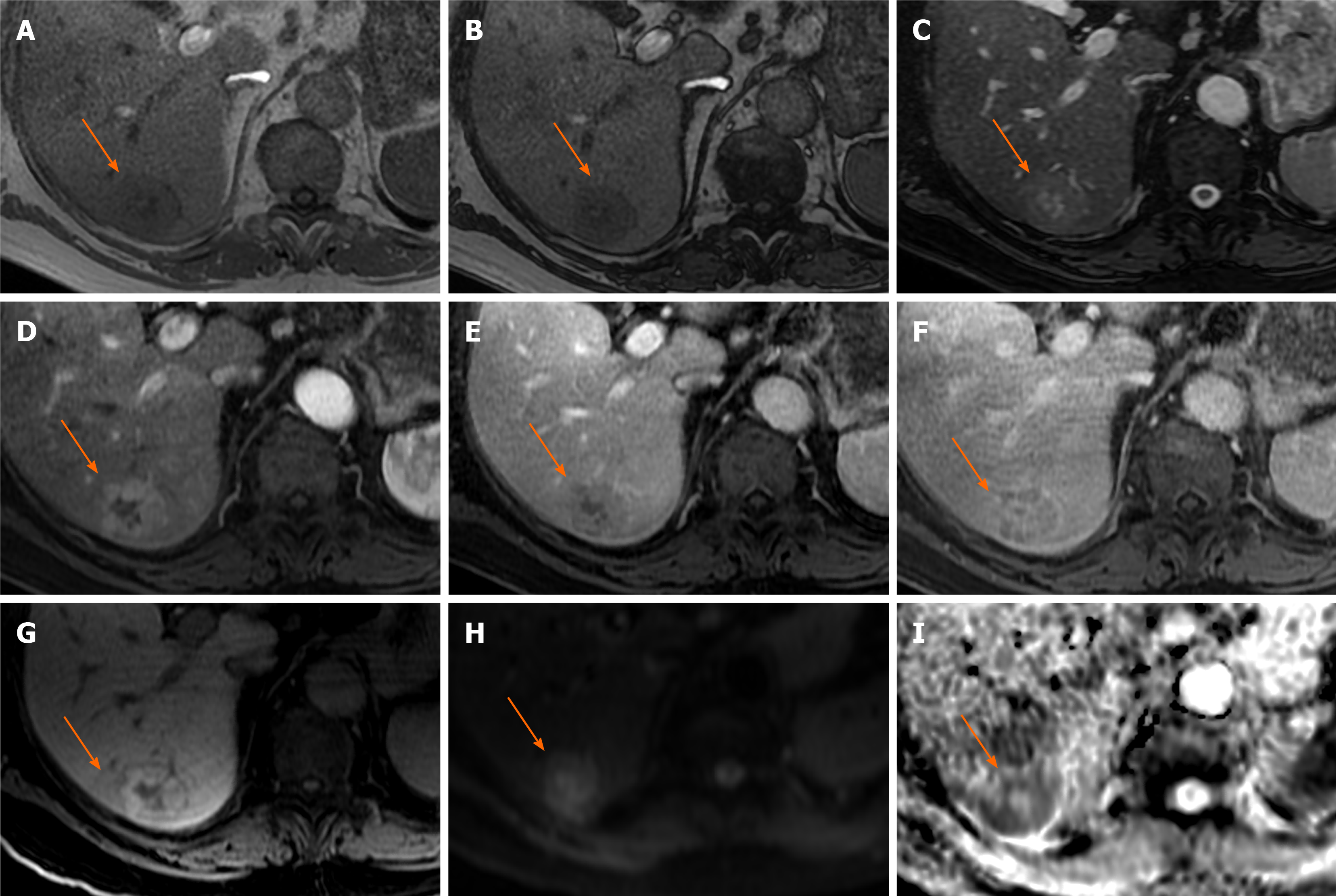
Figure 5 Hepatocellular carcinoma.
A 74-year-old man presented with incidental liver lesion found on routine computed tomography colonography. He had normal liver function and alpha-fetoprotein levels. The lesion had undergone further characterisation with magnetic resonance. A and B: There is no evidence of intralesional fat on T1-weighted in-phase (A) and out-of-phase (B) sequences; C: On T2-weighted images, the lesion is nearly isointense to the background liver and shows a hyperintense central scar, which can sometimes be seen in focal nodular hyperplasia; D-F: The lesion then demonstrates enhancement on the arterial phase (D) with evidence of washout as compared to background liver parenchyma on the portal venous (E) and delayed phases (F); there is also subtle peripheral enhancement on the delayed phase, likely representing a capsule, but the central scar remains largely unenhanced throughout; G: Hepatobiliary phase sequence demonstrates uptake of contrast in the majority of the lesion, with no uptake in the central scar and rim; H and I: diffusion-weighted imaging 500 (H) and low apparent diffusion coefficient (I) images suggest areas of diffusion restriction. Due to patient’s age, gender and indeterminate contrast characteristic, the lesion was resected. Histology showed the lesion was a well to moderately differentiated hepatocellular carcinoma. There was no background cirrhosis, but evidence of mild steatosis.
- Citation: Noreikaite J, Albasha D, Chidambaram V, Arora A, Katti A. Indeterminate liver lesions on gadoxetic acid-enhanced magnetic resonance imaging of the liver: Case-based radiologic-pathologic review. World J Hepatol 2021; 13(9): 1079-1097
- URL: https://www.wjgnet.com/1948-5182/full/v13/i9/1079.htm
- DOI: https://dx.doi.org/10.4254/wjh.v13.i9.1079


