Copyright
©The Author(s) 2021.
World J Hepatol. Sep 27, 2021; 13(9): 1079-1097
Published online Sep 27, 2021. doi: 10.4254/wjh.v13.i9.1079
Published online Sep 27, 2021. doi: 10.4254/wjh.v13.i9.1079
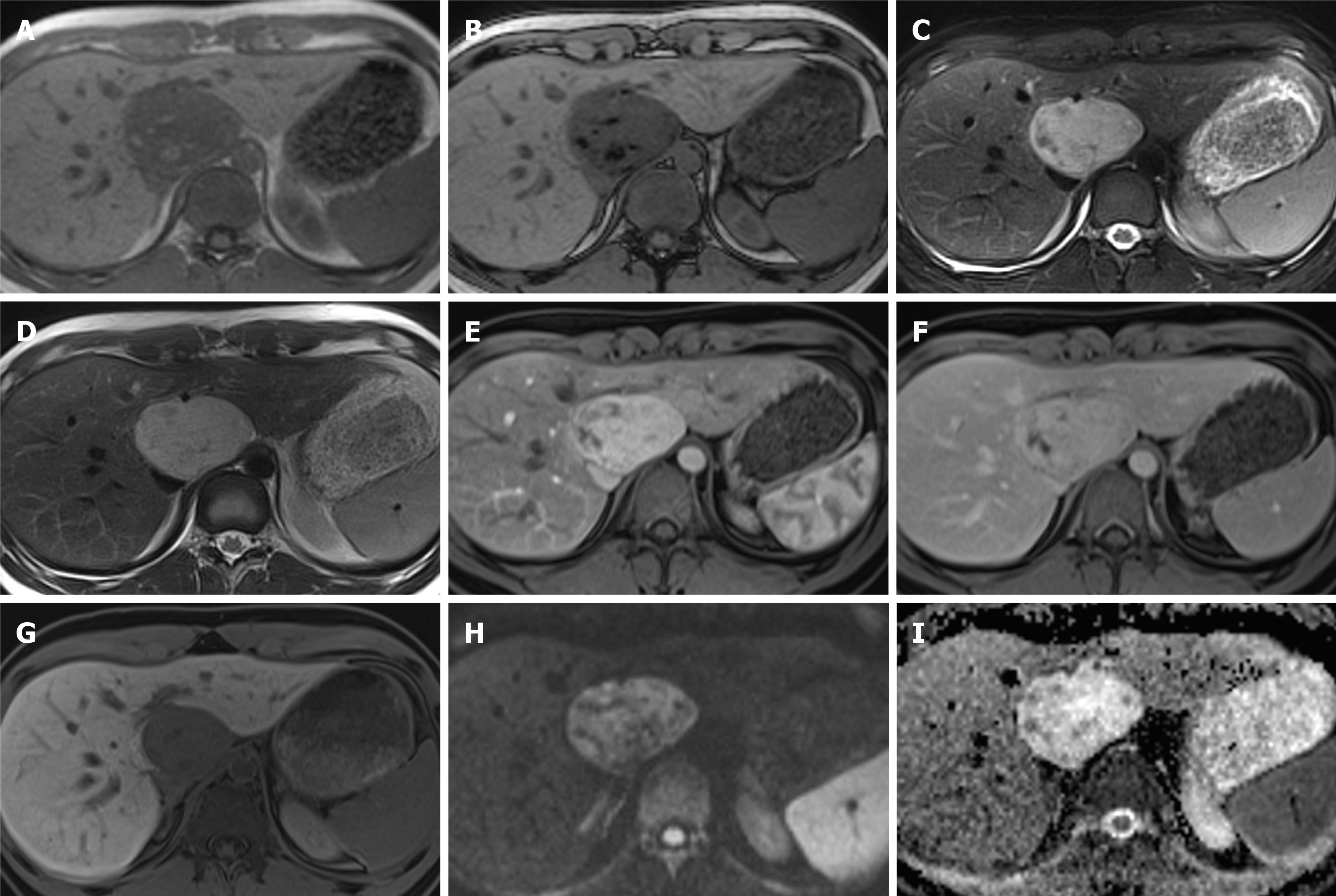
Figure 4 Hepatic angiomyolipoma.
A 21-year-old man referred by general practitioner for ultrasound of liver due to 6-mo history of intermittent abdominal pain and isolated raised bilirubin, treated as Gilbert’s syndrome. The patient had no prior medical history, no use of drugs or steroids and was not a heavy drinker. Incidental liver lesion was found and patient underwent subsequent magnetic resonance (MR) with gadoxetic acid to characterise this further. This was initially described as adenoma, but as the lesion increased in size on follow up imaging it was resected. Histology showed this to be an angiomyolipoma. A and B: MR demonstrates well-defined lesion with high signal foci on T1 in-phase (A) showing loss of signal on out-of-phase imaging (B); C and D: There are also hypointense foci on fat suppressed T2-weighted (C) when compared to T2-weighted imaging without fat suppression (D); E and F: The lesion shows enhancement on arterial phase (E) with no washout on equilibrium phase (F) and no pseudocapsule; G: There is no contrast uptake on hepatobiliary phase; H and I: No diffusion restriction as seen on diffusion-weighted imaging (H) and apparent diffusion coefficient (I) sequences.
- Citation: Noreikaite J, Albasha D, Chidambaram V, Arora A, Katti A. Indeterminate liver lesions on gadoxetic acid-enhanced magnetic resonance imaging of the liver: Case-based radiologic-pathologic review. World J Hepatol 2021; 13(9): 1079-1097
- URL: https://www.wjgnet.com/1948-5182/full/v13/i9/1079.htm
- DOI: https://dx.doi.org/10.4254/wjh.v13.i9.1079


