Copyright
©The Author(s) 2021.
World J Hepatol. Sep 27, 2021; 13(9): 1079-1097
Published online Sep 27, 2021. doi: 10.4254/wjh.v13.i9.1079
Published online Sep 27, 2021. doi: 10.4254/wjh.v13.i9.1079
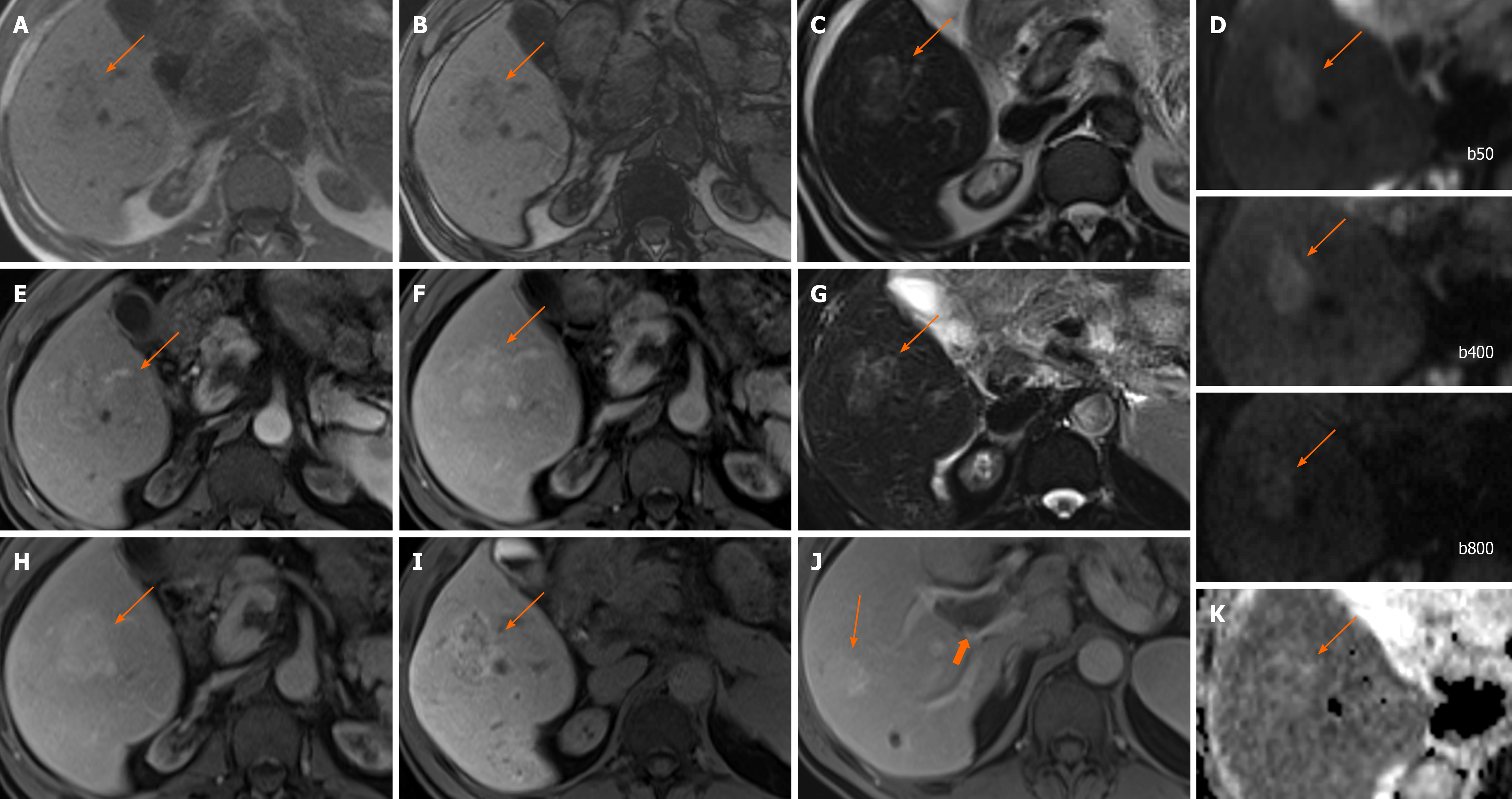
Figure 3 Focal nodular hyperplasia.
A 53-year-old woman with background of renal failure with renal transplant and history of autoimmune hepatitis since childhood. She underwent ultrasound (US) of the abdomen after an episode of pancreatitis which identified portal vein thrombosis. Subsequent unenhanced computed tomography (due to poor renal function) demonstrated a liver lesion in segment 5. Initially contrast US was attempted due to renal failure, which showed liver lesions to be multiple, but the lesions were indeterminate and subsequent magnetic resonance with gadoxetic acid was performed. Largest lesion in segment 5 selected as example. A and B: In-(A) and out-(B) of phase imaging shows some signal loss and mildly hypointense T1-weighted signal of the ill-defined right lobe lesion; C and G: T2-weighted without (C) and with fat suppression (G) show mildly hyperintense T2 signal; D and K: Diffusion-weighted imaging (D) and apparent diffusion coefficient (K) images show no diffusion restriction. E, F, and H: There is heterogenous enhancement on arterial phase (E) with no washout and slightly more homogenous contrast enhancement on portal venous (F) and delayed (H) phases; I and J: Heterogenous contrast uptake persists on hepatobiliary phase (I), which is mostly rim-like. Further similar lesion demonstrated on portal venous phase (J) in segment 7 (long arrow) and the known portal vein thrombus (short arrow). Initial radiological diagnosis favoured hepatocellular carcinoma. Liver function tests were normal. Initial non targeted liver biopsy was inconclusive for underlying cirrhosis. Second targeted lesion biopsy was performed. Both specimens were further reviewed in a national liver centre. Histology of the lesion was consistent with focal nodular hyperplasia and background liver demonstrated no cirrhosis, but signs consistent with nodular regenerative hyperplasia.
- Citation: Noreikaite J, Albasha D, Chidambaram V, Arora A, Katti A. Indeterminate liver lesions on gadoxetic acid-enhanced magnetic resonance imaging of the liver: Case-based radiologic-pathologic review. World J Hepatol 2021; 13(9): 1079-1097
- URL: https://www.wjgnet.com/1948-5182/full/v13/i9/1079.htm
- DOI: https://dx.doi.org/10.4254/wjh.v13.i9.1079


