Copyright
©The Author(s) 2021.
World J Hepatol. Sep 27, 2021; 13(9): 1079-1097
Published online Sep 27, 2021. doi: 10.4254/wjh.v13.i9.1079
Published online Sep 27, 2021. doi: 10.4254/wjh.v13.i9.1079
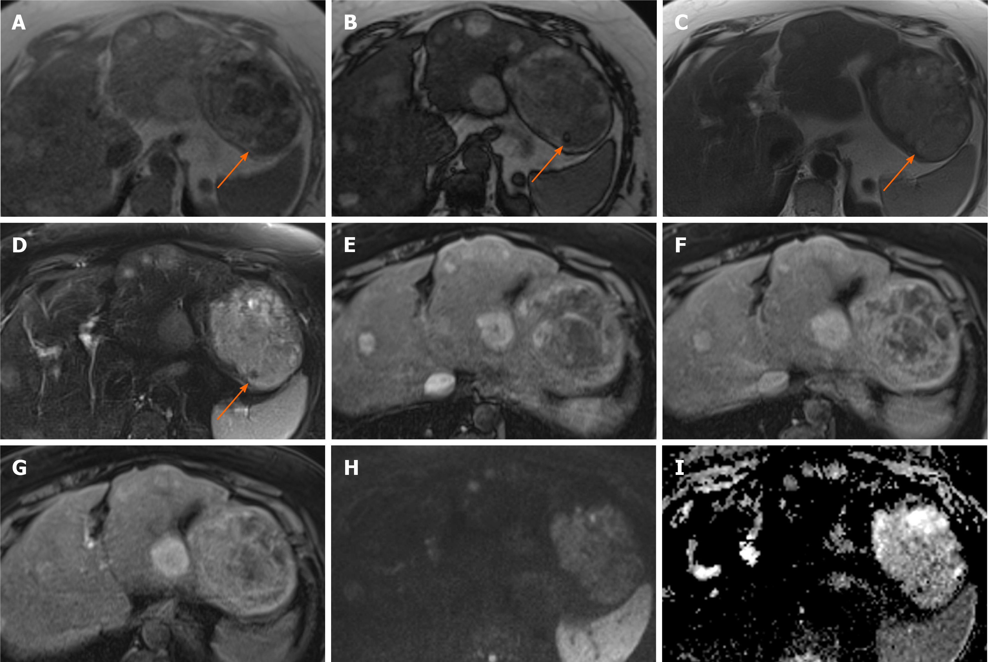
Figure 1 Hepatocellular adenoma.
A 42-year-old lady with congenital absence of portal vein and history of use of oral contraceptive medication presented with worsening jaundice. She underwent computed tomography that demonstrated multiple liver lesions that could not be characterised and subsequent magnetic resonance with gadoxetic acid was performed. This demonstrates multiple small lesions showing characteristics those of focal nodular hyperplasia. There is a further exophytic large lesion arising from the left liver lobe. The lesion is well-defined, T2 hyperintense and shows intratumoral fat (arrowed). A: In phase T1; B: Out-of-phase T1; C: T2-weighted imaging (T2-WI); D: Fat suppressed T2-WI; E-G: The arterial (E) and equilibrium (F) phase sequences demonstrates heterogenous enhancement with progressive filling in and there is contrast retention on hepatobiliary phase (G); H and I: Diffusion-weighted imaging (H) and apparent diffusion coefficient (I) sequences show no restricted diffusion. Due to atypical appearances this was resected and histology revealed this to be an adenoma with background steatotic liver.
- Citation: Noreikaite J, Albasha D, Chidambaram V, Arora A, Katti A. Indeterminate liver lesions on gadoxetic acid-enhanced magnetic resonance imaging of the liver: Case-based radiologic-pathologic review. World J Hepatol 2021; 13(9): 1079-1097
- URL: https://www.wjgnet.com/1948-5182/full/v13/i9/1079.htm
- DOI: https://dx.doi.org/10.4254/wjh.v13.i9.1079


