Copyright
©The Author(s) 2021.
World J Hepatol. Jun 27, 2021; 13(6): 699-708
Published online Jun 27, 2021. doi: 10.4254/wjh.v13.i6.699
Published online Jun 27, 2021. doi: 10.4254/wjh.v13.i6.699
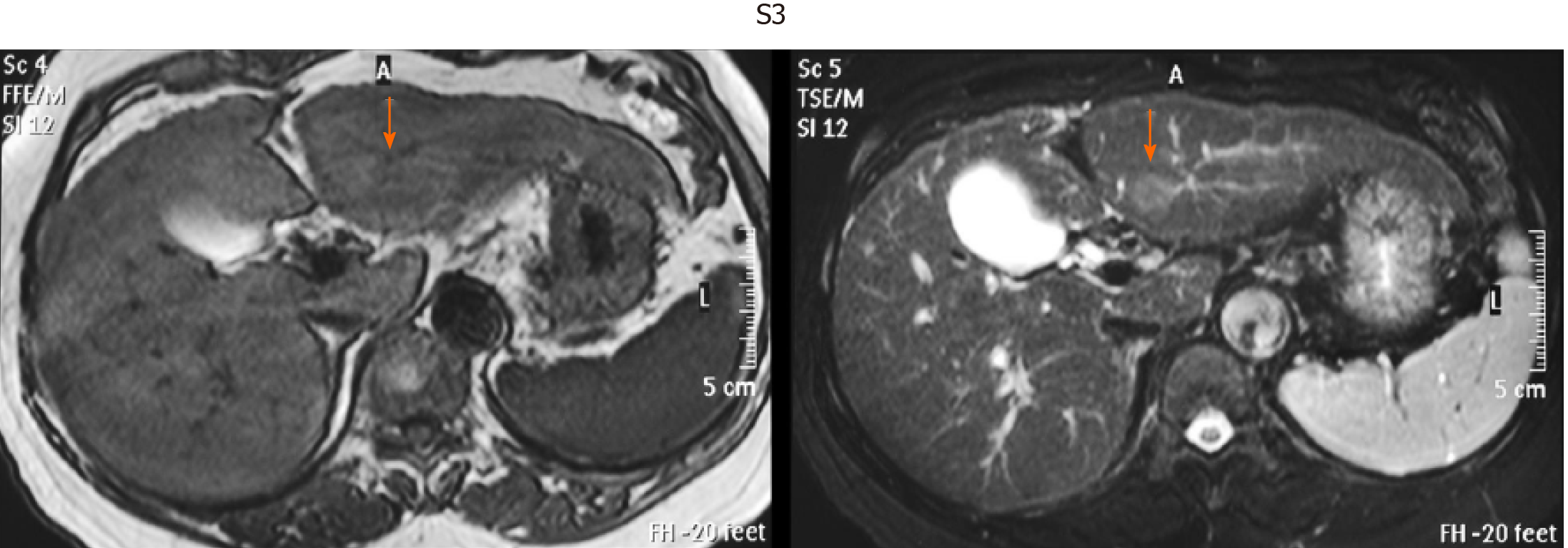
Figure 4 Representative image of very small hepatocellular carcinoma by unenhanced magnetic resonance imaging.
Hepatocellular carcinoma in S3 segment. T1-weighted image (left, light dark spot). T2-weighted image (right, light white spot).
- Citation: Tarao K, Nozaki A, Komatsu H, Komatsu T, Taguri M, Tanaka K, Yoshida T, Koyasu H, Chuma M, Numata K, Maeda S. Comparison of unenhanced magnetic resonance imaging and ultrasound in detecting very small hepatocellular carcinoma. World J Hepatol 2021; 13(6): 699-708
- URL: https://www.wjgnet.com/1948-5182/full/v13/i6/699.htm
- DOI: https://dx.doi.org/10.4254/wjh.v13.i6.699


