Copyright
©The Author(s) 2021.
World J Hepatol. Jun 27, 2021; 13(6): 650-661
Published online Jun 27, 2021. doi: 10.4254/wjh.v13.i6.650
Published online Jun 27, 2021. doi: 10.4254/wjh.v13.i6.650
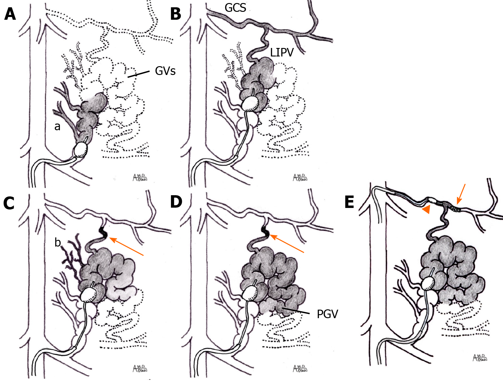
Figure 2 Illustration of the balloon-occluded retrograde transvenous obliteration procedure.
A: Balloon-occluded retrograde transvenous venography (BRTV). The initial BRTV does not visualize the main body of the gastric varices (GVs) because multiple draining vessels are present (a); B: When the balloon catheter is advanced beyond the small drainage vessels (downgrading method), the relatively large diameter left inferior phrenic vein (LIPV) becomes visualized as another drainage route to the gastrocaval shunt (GCS); C: GVs become visualized when selective coil embolization (arrow) of the LIPV is performed. As small amounts of sclerosant are injected sequentially over time, the smaller drainage vessels (b) are gradually embolized (stepwise injection method); D: After stepwise injection, BRTV demonstrated the GVs in their entirety as well as the inflowing posterior gastric vein; E: If selective coil embolization of the LIPV is impossible, the GCS should be occluded with another balloon catheter for balloon-occluded retrograde transvenous obliteration (BRTO) (dual-BRTO). Selective coil embolization of the LIPV branch (arrow) is performed through the catheter via the GCS. PGV: Posterior gastric vein; GVs: Gastric varices; LIPV: Left inferior phrenic vein; GCS: Gastrocaval shunt.
- Citation: Waguri N, Osaki A, Watanabe Y. Balloon-occluded retrograde transvenous obliteration for treatment of gastric varices. World J Hepatol 2021; 13(6): 650-661
- URL: https://www.wjgnet.com/1948-5182/full/v13/i6/650.htm
- DOI: https://dx.doi.org/10.4254/wjh.v13.i6.650


