Copyright
©The Author(s) 2021.
World J Hepatol. Apr 27, 2021; 13(4): 515-521
Published online Apr 27, 2021. doi: 10.4254/wjh.v13.i4.515
Published online Apr 27, 2021. doi: 10.4254/wjh.v13.i4.515
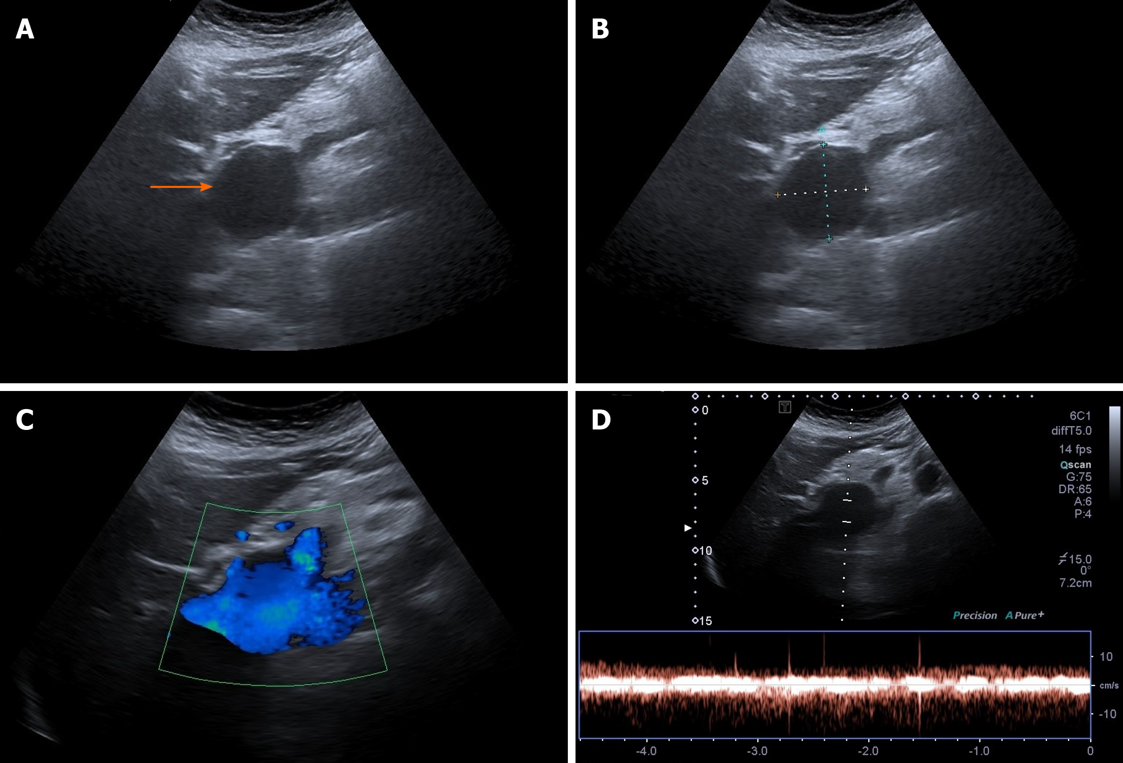
Figure 2 Abdominal ultrasonographic imaging of Case 2.
A: Extrahepatic anechoic saccular lesion, indicating an aneurysmal dilation of the extrahepatic portal vein (arrow); B: The anechoic lesion was 42.3 mm at its maximal diameter; C: Colour Doppler showed hepatopetal venous flow in the extrahepatic aneurysmal dilated vessel; D: Doppler recording showed pulsating flow of venous type.
- Citation: Priadko K, Romano M, Vitale LM, Niosi M, De Sio I. Asymptomatic portal vein aneurysm: Three case reports. World J Hepatol 2021; 13(4): 515-521
- URL: https://www.wjgnet.com/1948-5182/full/v13/i4/515.htm
- DOI: https://dx.doi.org/10.4254/wjh.v13.i4.515


