Copyright
©The Author(s) 2021.
World J Hepatol. Apr 27, 2021; 13(4): 515-521
Published online Apr 27, 2021. doi: 10.4254/wjh.v13.i4.515
Published online Apr 27, 2021. doi: 10.4254/wjh.v13.i4.515
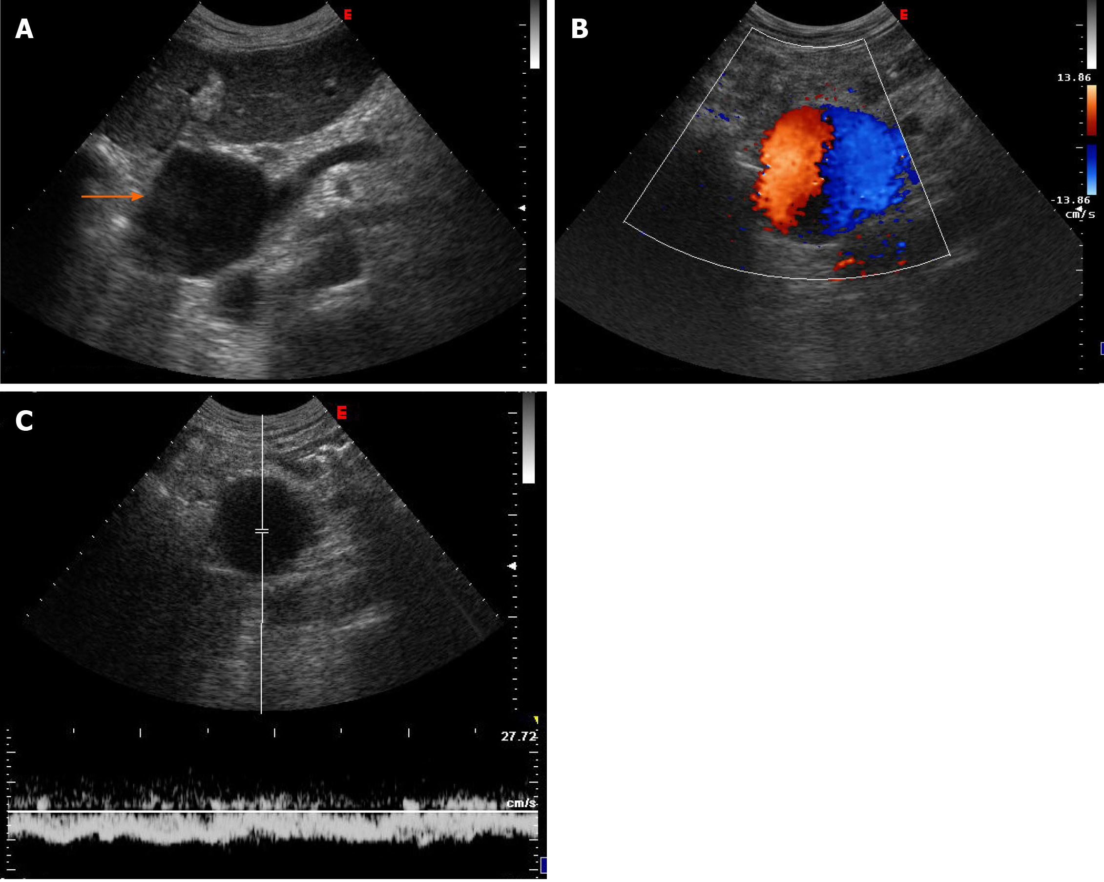
Figure 1 Abdominal ultrasonographic imaging of Case 1.
A: Anechoic lesion corresponding to a notably dilated extrahepatic portal vein (arrow); B: Colour Doppler showed the “Korean flag” pathological sign in the dilated portal vein; C: Doppler recording showed flat venous flow.
- Citation: Priadko K, Romano M, Vitale LM, Niosi M, De Sio I. Asymptomatic portal vein aneurysm: Three case reports. World J Hepatol 2021; 13(4): 515-521
- URL: https://www.wjgnet.com/1948-5182/full/v13/i4/515.htm
- DOI: https://dx.doi.org/10.4254/wjh.v13.i4.515


