Copyright
©The Author(s) 2021.
World J Hepatol. Mar 27, 2021; 13(3): 300-314
Published online Mar 27, 2021. doi: 10.4254/wjh.v13.i3.300
Published online Mar 27, 2021. doi: 10.4254/wjh.v13.i3.300
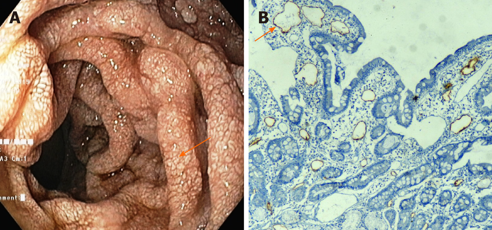
Figure 4 Intestinal lymphangiectasia in a patient with cirrhosis.
A: Upper gastrointestinal endoscopy of a patient showing whitish swollen villi in the duodenum, suggestive of intestinal lymphangiectasia; B: On immunohistochemistry (× 10), markedly dilated vessels were seen in the lamina which showed strong D2-40 positivity indicating dilated lymphatics.
- Citation: Kumar R, Anand U, Priyadarshi RN. Lymphatic dysfunction in advanced cirrhosis: Contextual perspective and clinical implications. World J Hepatol 2021; 13(3): 300-314
- URL: https://www.wjgnet.com/1948-5182/full/v13/i3/300.htm
- DOI: https://dx.doi.org/10.4254/wjh.v13.i3.300


