Copyright
©The Author(s) 2021.
World J Hepatol. Dec 27, 2021; 13(12): 2192-2200
Published online Dec 27, 2021. doi: 10.4254/wjh.v13.i12.2192
Published online Dec 27, 2021. doi: 10.4254/wjh.v13.i12.2192
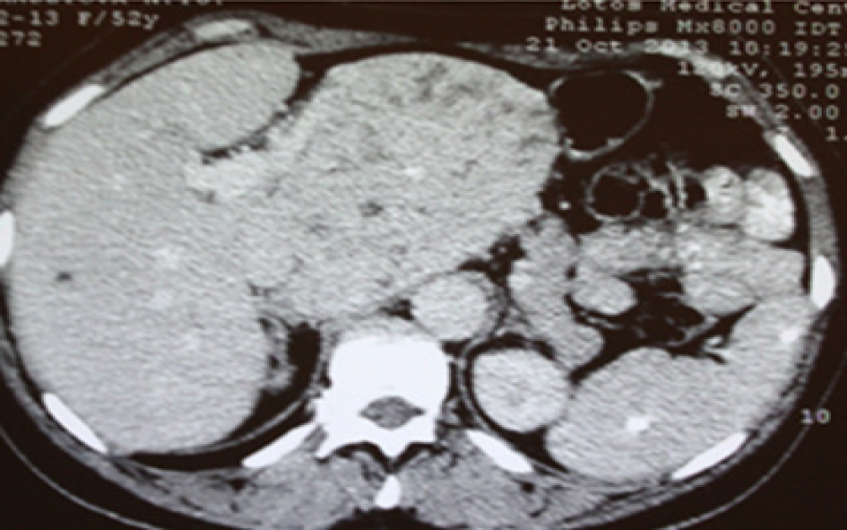
Figure 2 Abdominal computed tomography with contrast enhancement: Tumor invades segment I of the liver (longitudinal section).
Ill-defined contrast-enhancing, multilobulated cystic lesion involving segments II, III, VI and VIII.
- Citation: Kovalenko YA, Zharikov YO, Kiseleva YV, Goncharov AB, Shevchenko TV, Gurmikov BN, Kalinin DV, Zhao AV. Rare primary mature teratoma of the liver: A case report. World J Hepatol 2021; 13(12): 2192-2200
- URL: https://www.wjgnet.com/1948-5182/full/v13/i12/2192.htm
- DOI: https://dx.doi.org/10.4254/wjh.v13.i12.2192


