Copyright
©The Author(s) 2021.
World J Hepatol. Dec 27, 2021; 13(12): 1936-1955
Published online Dec 27, 2021. doi: 10.4254/wjh.v13.i12.1936
Published online Dec 27, 2021. doi: 10.4254/wjh.v13.i12.1936
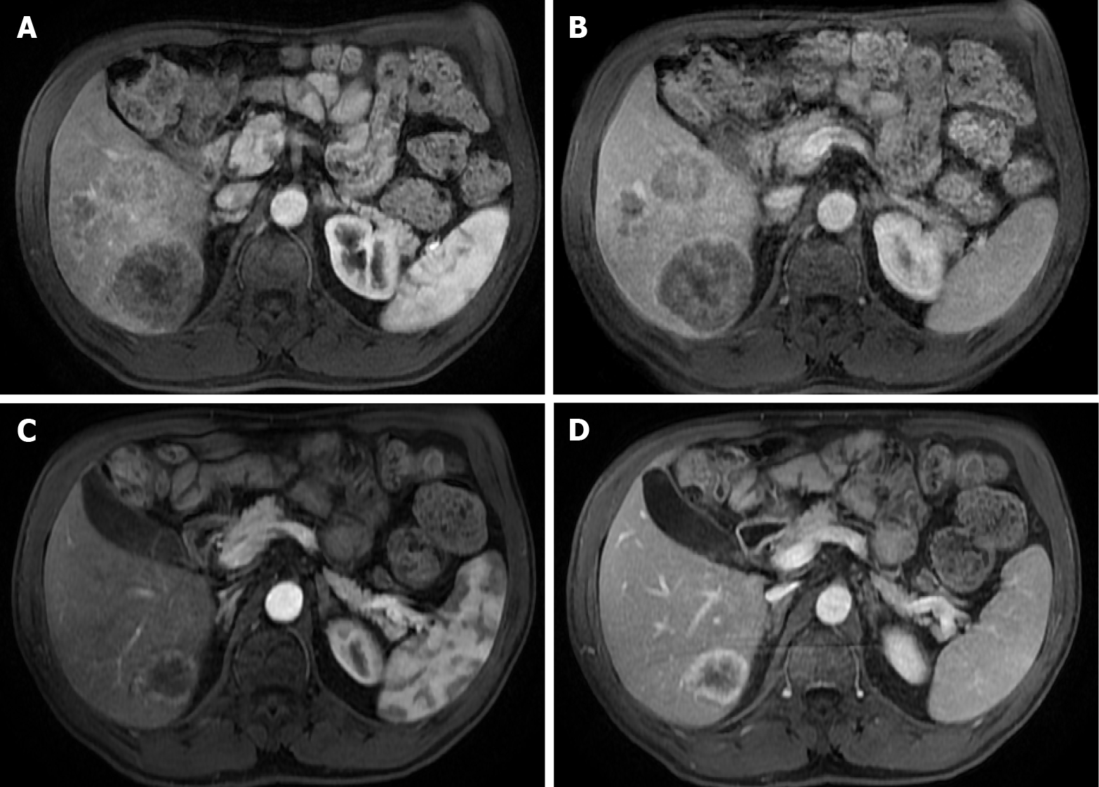
Figure 13 A 71-year-old man with unresectable CRLM.
A and C: Axial fat saturated (FS) contrast-enhanced magnetic resonance imaging (CE-MRI) T1-weighted imaging (WI) in the arterial phase; B and D: Axial FS CE-MRI T1-WI in the portal-venous phase. Initial presentation of three heterogeneous hepatic lesions corresponding to unresectable CRLM before treatment (A and B). After chemotherapy (C and D), the patient presented partial response, with the disappearance of two lesions and reduced size of the larger lesion, which still presents viable peripheral tumor.
- Citation: Freitas PS, Janicas C, Veiga J, Matos AP, Herédia V, Ramalho M. Imaging evaluation of the liver in oncology patients: A comparison of techniques. World J Hepatol 2021; 13(12): 1936-1955
- URL: https://www.wjgnet.com/1948-5182/full/v13/i12/1936.htm
- DOI: https://dx.doi.org/10.4254/wjh.v13.i12.1936


