Copyright
©The Author(s) 2021.
World J Hepatol. Dec 27, 2021; 13(12): 1936-1955
Published online Dec 27, 2021. doi: 10.4254/wjh.v13.i12.1936
Published online Dec 27, 2021. doi: 10.4254/wjh.v13.i12.1936
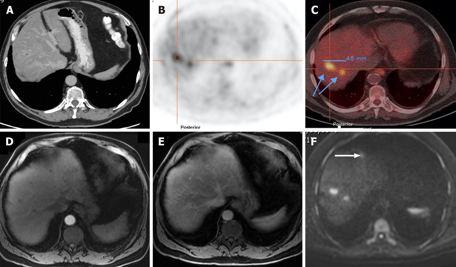
Figure 12 A 71-year-old man with colorectal carcinoma presenting with liver metastases.
A: Axial contrast-enhanced computed tomography (CE-CT) in the portal-venous phase shows barely identified non-specific liver micronodules; B and C: Fluorodeoxyglucose positron emission tomography (PET)-CT shows two hypermetabolic lesions in the right lobe, consistent with viable neoplastic tissue; D: Axial fat saturated (FS) CE- magnetic resonance imaging (MRI) T1-weighted imaging (WI) in the arterial phase shows hypovascular liver lesions; E: Axial FS CE-MRI T1-WI in the venous phase confirms the liver metastases, showing hypointense nodules with venous ring enhancement; F: In the diffusion-weighted imaging study, these lesions are more conspicuous. Also, an additional metastasis (arrow) that was not detected either by CE-CT, CE-MRI, or PET-CT is shown.
- Citation: Freitas PS, Janicas C, Veiga J, Matos AP, Herédia V, Ramalho M. Imaging evaluation of the liver in oncology patients: A comparison of techniques. World J Hepatol 2021; 13(12): 1936-1955
- URL: https://www.wjgnet.com/1948-5182/full/v13/i12/1936.htm
- DOI: https://dx.doi.org/10.4254/wjh.v13.i12.1936


