Copyright
©The Author(s) 2021.
World J Hepatol. Dec 27, 2021; 13(12): 1936-1955
Published online Dec 27, 2021. doi: 10.4254/wjh.v13.i12.1936
Published online Dec 27, 2021. doi: 10.4254/wjh.v13.i12.1936
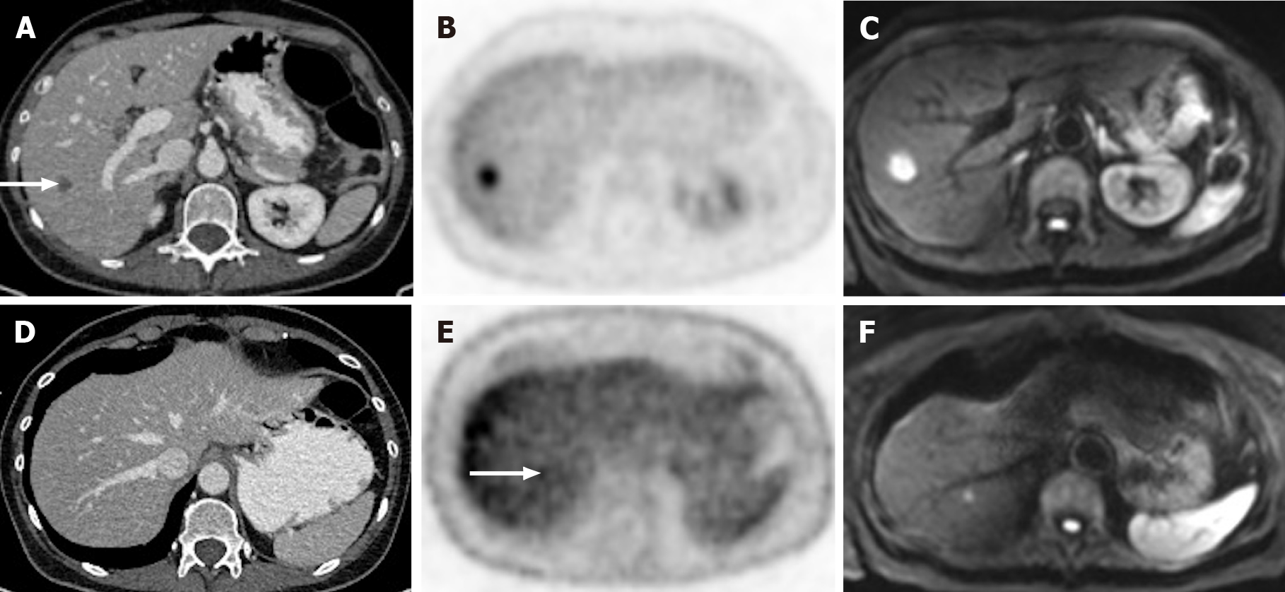
Figure 11 A 65-year-old woman with colorectal carcinoma shows liver metastasis in segment VII.
A: Axial contrast-enhanced computed tomography (CE-CT) reveals a hypodense lesion corresponding to liver metastasis (arrow); B: Fluorodeoxyglucose (FDG) positron emission tomography (PET)-CT confirms metastatic origin; D: Axial CE-CT shows no apparent lesion; E: FDG PET-CT shows an additional barely visible nodule not seen in CT (arrow); C and F: Diffusion-weighted imaging confirmed that both lesions were secondary.
- Citation: Freitas PS, Janicas C, Veiga J, Matos AP, Herédia V, Ramalho M. Imaging evaluation of the liver in oncology patients: A comparison of techniques. World J Hepatol 2021; 13(12): 1936-1955
- URL: https://www.wjgnet.com/1948-5182/full/v13/i12/1936.htm
- DOI: https://dx.doi.org/10.4254/wjh.v13.i12.1936


