Copyright
©The Author(s) 2021.
World J Hepatol. Dec 27, 2021; 13(12): 1936-1955
Published online Dec 27, 2021. doi: 10.4254/wjh.v13.i12.1936
Published online Dec 27, 2021. doi: 10.4254/wjh.v13.i12.1936
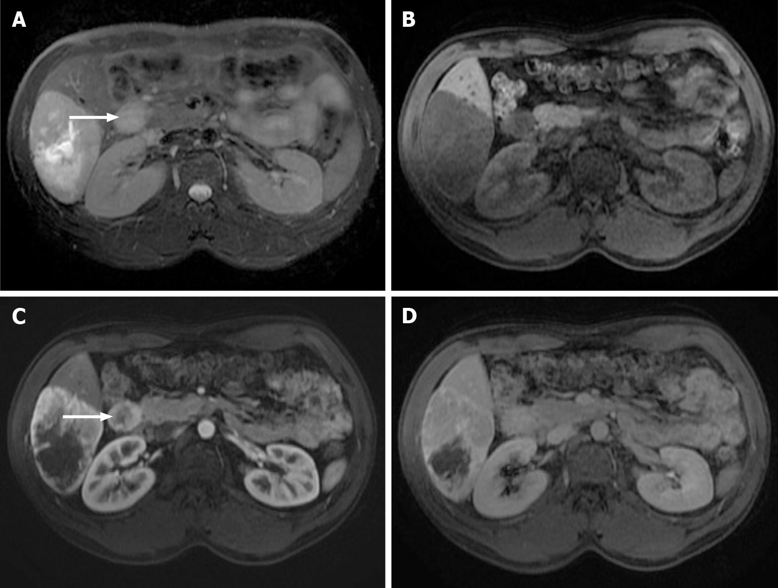
Figure 9 Images show a large liver metastasis from a duodenal neuroendocrine tumor.
A: In the axial fat saturated (FS) T2-weighted imaging (WI), the liver metastasis is characterized by hyperintense central necrosis delimited by a lesser intense viable tumor. Note the duodenal neuroendocrine tumor (arrow); B: Axial FS non-contrast-enhanced magnetic resonance imaging (CE-MRI) T1-WI shows large hypointense liver metastasis; C: Axial FS CE-MRI T1-WI in the arterial phase demonstrates viable tumor with avid heterogeneous enhancement. The primary lesion is also hypervascular and depicted in the 2nd portion of the duodenum (arrow); D: Axial FS CE-MRI T1-WI in the interstitial phase reveals fading of the lesion.
- Citation: Freitas PS, Janicas C, Veiga J, Matos AP, Herédia V, Ramalho M. Imaging evaluation of the liver in oncology patients: A comparison of techniques. World J Hepatol 2021; 13(12): 1936-1955
- URL: https://www.wjgnet.com/1948-5182/full/v13/i12/1936.htm
- DOI: https://dx.doi.org/10.4254/wjh.v13.i12.1936


