Copyright
©The Author(s) 2021.
World J Hepatol. Dec 27, 2021; 13(12): 1936-1955
Published online Dec 27, 2021. doi: 10.4254/wjh.v13.i12.1936
Published online Dec 27, 2021. doi: 10.4254/wjh.v13.i12.1936
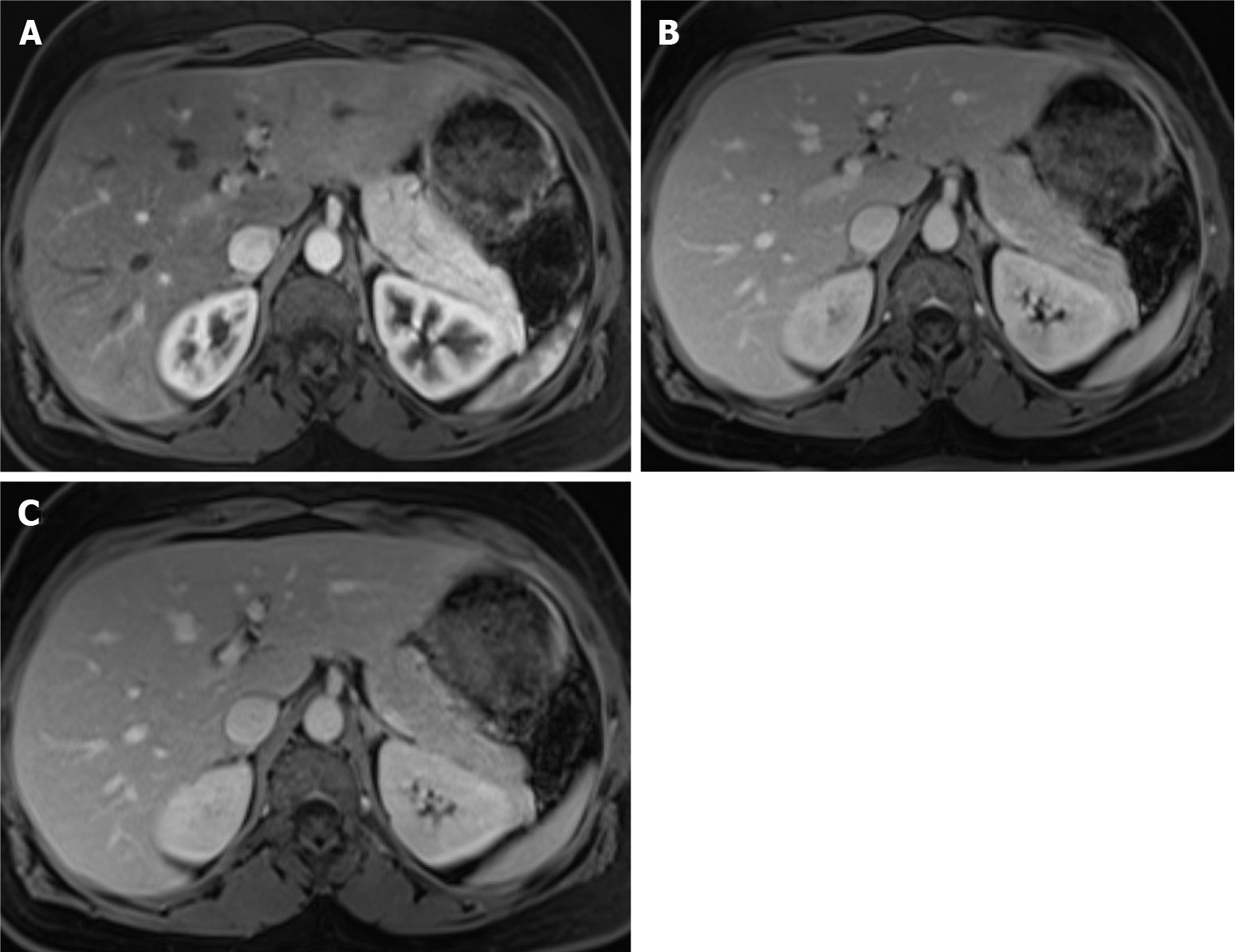
Figure 2 Dynamic phases of enhancement.
A: Late hepatic arterial phase. It is characterized by contrast in hepatic arteries and portal veins, not in hepatic veins. It is helpful for hypervascular lesions and perfusional abnormalities. Note that the normal pancreas enhances greater than the liver; B: Portal venous phase. It is recognized by the contrast in the hepatic and portal veins. It is useful mainly for hypovascular lesion detection; C: Interstitial or delayed phase. It is helpful for lesion characterization, especially for late enhancement perception.
- Citation: Freitas PS, Janicas C, Veiga J, Matos AP, Herédia V, Ramalho M. Imaging evaluation of the liver in oncology patients: A comparison of techniques. World J Hepatol 2021; 13(12): 1936-1955
- URL: https://www.wjgnet.com/1948-5182/full/v13/i12/1936.htm
- DOI: https://dx.doi.org/10.4254/wjh.v13.i12.1936


