Copyright
©The Author(s) 2021.
World J Hepatol. Dec 27, 2021; 13(12): 1892-1908
Published online Dec 27, 2021. doi: 10.4254/wjh.v13.i12.1892
Published online Dec 27, 2021. doi: 10.4254/wjh.v13.i12.1892
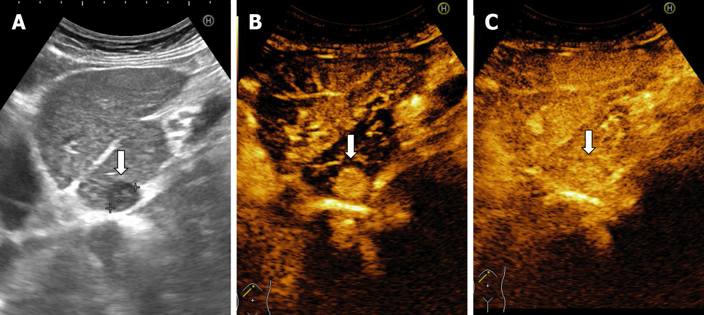
Figure 23 A difficult diagnosis in a case of a flash-filling hemangioma in a woman with hepatitis C liver cirrhosis.
A: On B mode ultrasound is observed inhomogeneous liver structure and enlargement of caudate lobe. A small hypoechoic lesion is detected in the caudate lobe; B: Flash-filling enhancement in arterial phase is noticed that is similar to the enhancement of an hepatocellular carcinoma; C: Even in the late phase the liver lesion had the same enhancement comparative with liver, the segmental resection was performed. On histopathological exam the conclusion was: liver hemangioma.
- Citation: Sandulescu LD, Urhut CM, Sandulescu SM, Ciurea AM, Cazacu SM, Iordache S. One stop shop approach for the diagnosis of liver hemangioma. World J Hepatol 2021; 13(12): 1892-1908
- URL: https://www.wjgnet.com/1948-5182/full/v13/i12/1892.htm
- DOI: https://dx.doi.org/10.4254/wjh.v13.i12.1892


