Copyright
©The Author(s) 2021.
World J Hepatol. Nov 27, 2021; 13(11): 1484-1493
Published online Nov 27, 2021. doi: 10.4254/wjh.v13.i11.1484
Published online Nov 27, 2021. doi: 10.4254/wjh.v13.i11.1484
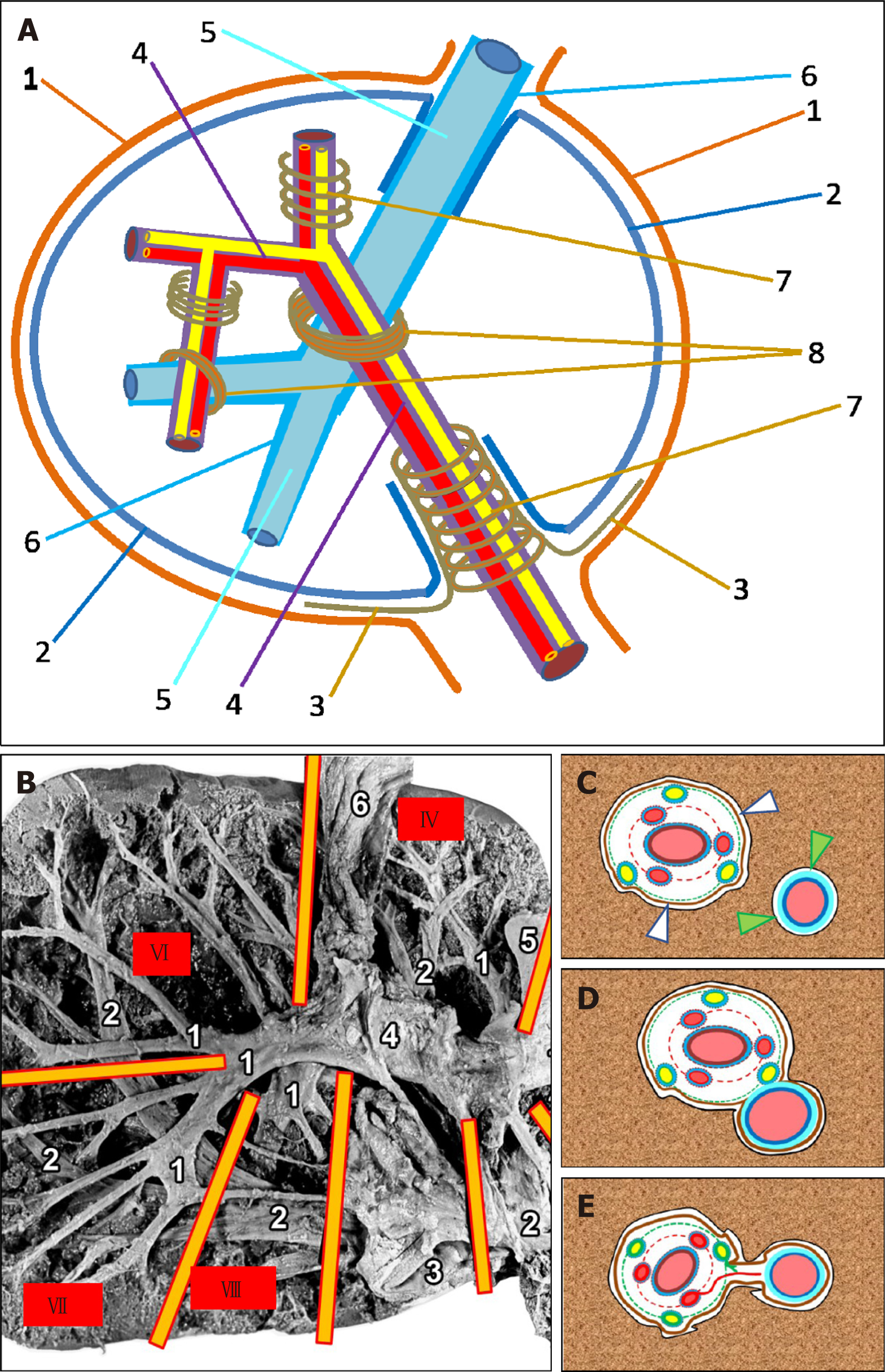
Figure 1 Connective tissue structures and their relationship in the liver.
A: 1: Peritoneum; 2: Liver capsule (Laennec's capsule); 3: Hilar plate (Walaeus vasculo-biliary sheath); 4: Portal tract; 5: Hepatic vein and its tributaries; 6: Connective tissue sheath of a hepatic vein; 7: Portal tract surrounded by Glisson's capsule (Glissonean pedicle); 8: Porta-caval fibrous connection (PCFC); Arrowhead: the fissure among the Laennec's capsule (proper hepatic capsule, PHC) and the Glisson's capsule; B: Intrahepatic portal tracts and hepatic veins of the human liver after maceration from the visceral surface (preparation from the private archive of Professor Chanukvadze I); Intersection of portal tracts and the hepatic veins. Yellow lines show the borders among the liver segments enumeration of which is shown in red quadrats. 1: Portal tract; 2: Hepatic veins and their tributaries; 3: Inferior vena cava; 4: Walaeus vasculo-biliary sheath; 5: Round ligament; 6: Gallbladder; C: Section of liver tissue containing the portal tract and hepatic vein (scheme). White arrowhead: the fissure among the Laennec's capsule (PHC) and the Glisson's capsule; Green arrowhead: the fissure among the Laennec's capsule (PHC) and connective-tissue sheath surrounding the hepatic vein; D: Area of complete fusion of the Glisson's capsule and a connective-tissue sheath surrounding the hepatic vein (scheme); E: Plate-shaped PCFC (scheme).
- Citation: Patarashvili L, Gvidiani S, Azmaipharashvili E, Tsomaia K, Sareli M, Kordzaia D, Chanukvadze I. Porta-caval fibrous connections — the lesser-known structure of intrahepatic connective-tissue framework: A unified view of liver extracellular matrix. World J Hepatol 2021; 13(11): 1484-1493
- URL: https://www.wjgnet.com/1948-5182/full/v13/i11/1484.htm
- DOI: https://dx.doi.org/10.4254/wjh.v13.i11.1484


