Copyright
©The Author(s) 2021.
World J Hepatol. Oct 27, 2021; 13(10): 1316-1327
Published online Oct 27, 2021. doi: 10.4254/wjh.v13.i10.1316
Published online Oct 27, 2021. doi: 10.4254/wjh.v13.i10.1316
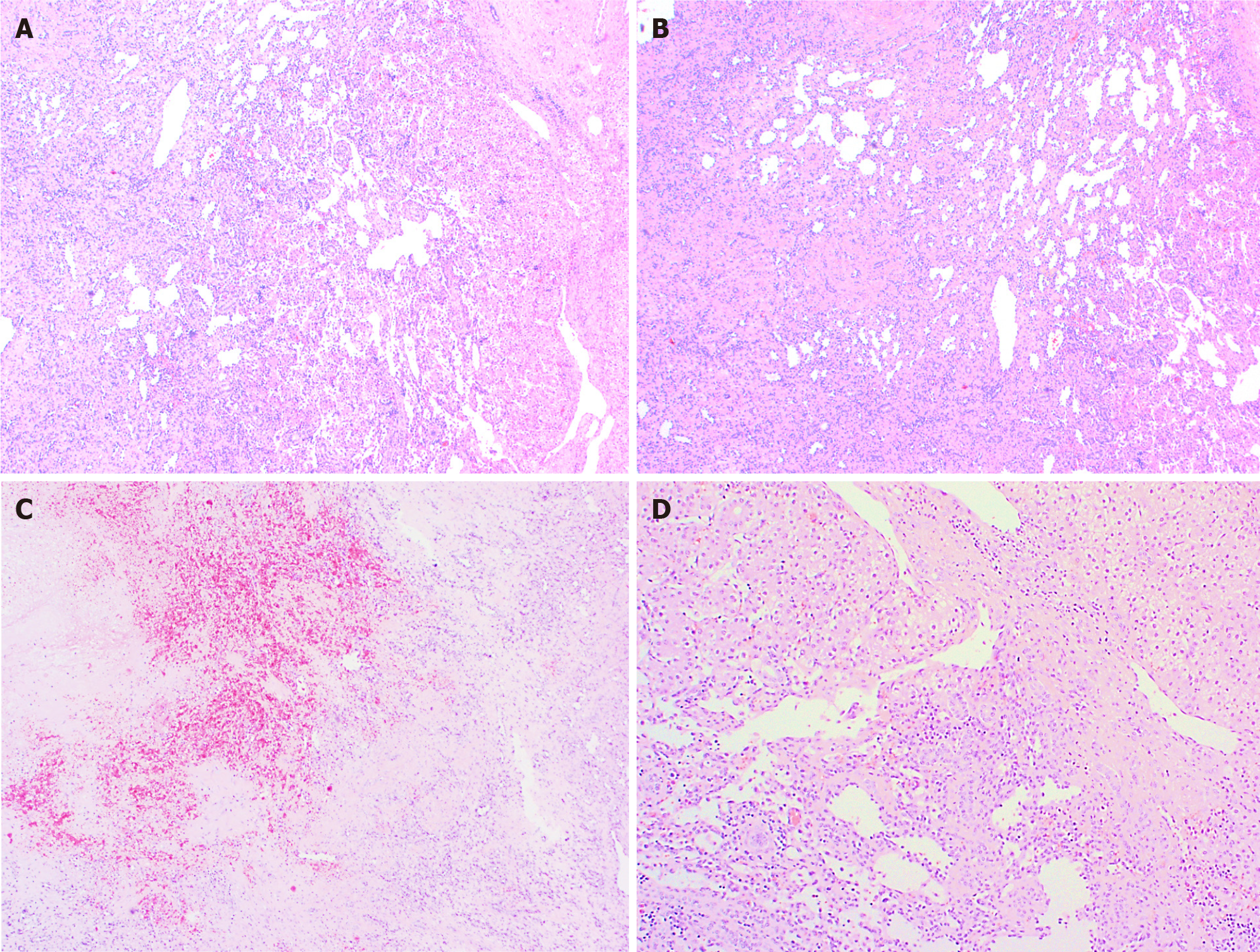
Figure 2 Hepatic congenital hemangioma.
A: A relatively well-demarcated vascular lesion; B: Lobules of variable sized, mostly small thin-walled vascular spaces and more abundant larger vessels at the periphery; C: Necrotic and hemorrhagic areas in the central part (area of involution); D: Entrapment of hepatocytes and bile ducts in interface areas.
- Citation: Cordier F, Hoorens A, Van Dorpe J, Creytens D. Pediatric vascular tumors of the liver: Review from the pathologist’s point of view. World J Hepatol 2021; 13(10): 1316-1327
- URL: https://www.wjgnet.com/1948-5182/full/v13/i10/1316.htm
- DOI: https://dx.doi.org/10.4254/wjh.v13.i10.1316


