Copyright
©The Author(s) 2020.
World J Hepatol. Jun 27, 2020; 12(6): 262-276
Published online Jun 27, 2020. doi: 10.4254/wjh.v12.i6.262
Published online Jun 27, 2020. doi: 10.4254/wjh.v12.i6.262
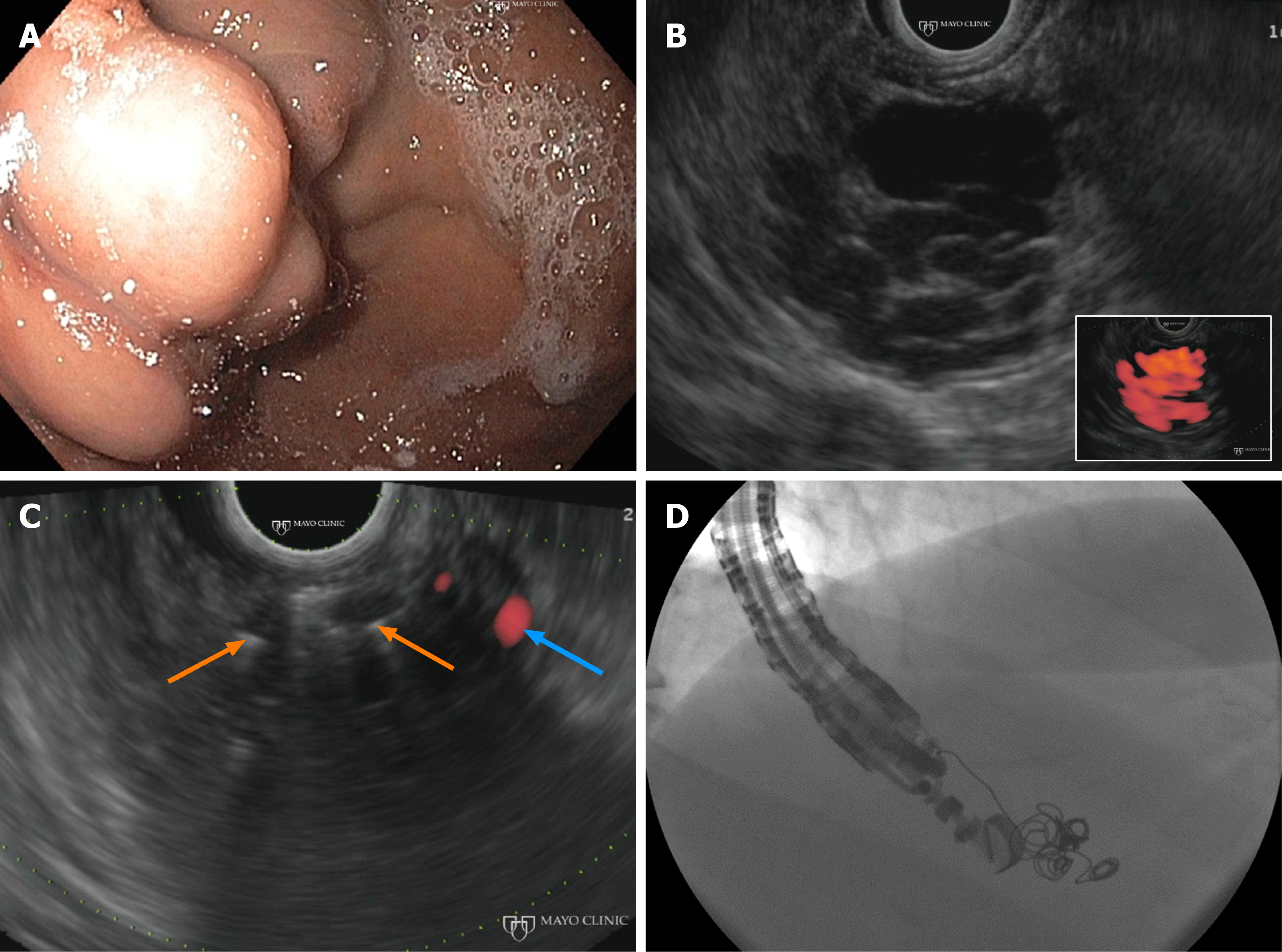
Figure 4 Endoscopic ultrasound-guided management of gastric varices.
A: Gastric varices seen on endoscopy. B: Gastric varices appear anechoic on endoscopic ultrasound (EUS) grey-scale and are highlighted red by Doppler study (inset). C: Injection of embolization coils (orange arrows) into the varices results in near complete resolution of blood flow (blue arrow). D: Fluoroscopic visualization of EUS-guided coil embolization.
- Citation: Fung BM, Abadir AP, Eskandari A, Levy MJ, Tabibian JH. Endoscopic ultrasound in chronic liver disease. World J Hepatol 2020; 12(6): 262-276
- URL: https://www.wjgnet.com/1948-5182/full/v12/i6/262.htm
- DOI: https://dx.doi.org/10.4254/wjh.v12.i6.262


