Copyright
©The Author(s) 2020.
World J Hepatol. May 27, 2020; 12(5): 207-219
Published online May 27, 2020. doi: 10.4254/wjh.v12.i5.207
Published online May 27, 2020. doi: 10.4254/wjh.v12.i5.207
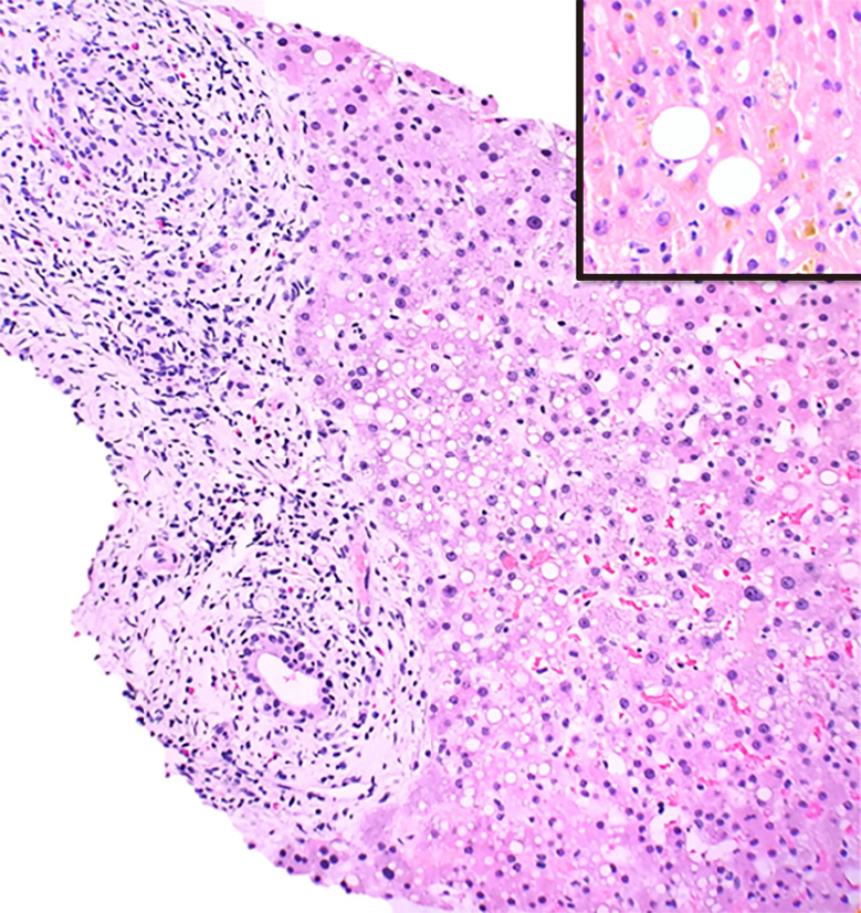
Figure 5 Liver injury secondary to hydroxycut.
Liver parenchyma with expansion of the portal areas by a mixed inflammatory infiltrate comprising lymphocytes, eosinophils, rare plasma cells and scattered neutrophils. There is evidence of bile duct injury with infiltration of the bile duct epithelium by inflammatory cells (20 ×). The lobules show macrovesicular steatosis, feathery degeneration of hepatocytes, minimal lobulitis, and focal cholestasis (insert 40 ×).
- Citation: Siddique AS, Siddique O, Einstein M, Urtasun-Sotil E, Ligato S. Drug and herbal/dietary supplements-induced liver injury: A tertiary care center experience. World J Hepatol 2020; 12(5): 207-219
- URL: https://www.wjgnet.com/1948-5182/full/v12/i5/207.htm
- DOI: https://dx.doi.org/10.4254/wjh.v12.i5.207


