Copyright
©The Author(s) 2020.
World J Hepatol. May 27, 2020; 12(5): 207-219
Published online May 27, 2020. doi: 10.4254/wjh.v12.i5.207
Published online May 27, 2020. doi: 10.4254/wjh.v12.i5.207
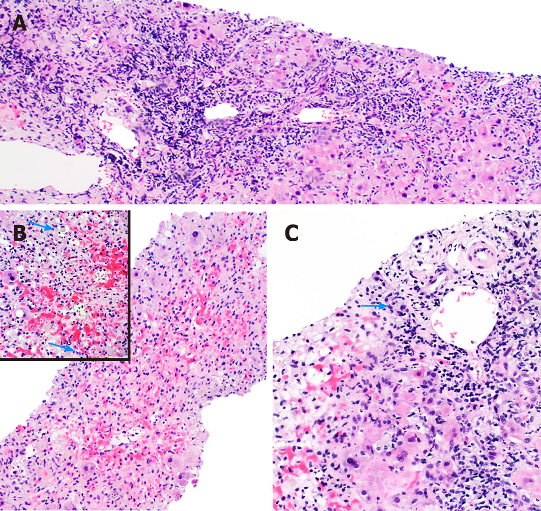
Figure 4 Liver injury secondary to C4 extreme.
A: The liver parenchyma demonstrates a dense mononuclear inflammatory infiltrate involving the portal tracts with severe interface necroinflammatory activity (10 ×); B: In the pericentral and midzonal area of the hepatic lobules (zone 2 and 3), there is extensive hemorrhagic necrosis with extravasation of red blood cells from the central vein into the sinusoidal spaces (10 ×). Hepatocellular and canalicular cholestasis is seen (arrow) (insert 40 ×); C: The portal areas display extensive bile duct damage (arrow) (20 ×).
- Citation: Siddique AS, Siddique O, Einstein M, Urtasun-Sotil E, Ligato S. Drug and herbal/dietary supplements-induced liver injury: A tertiary care center experience. World J Hepatol 2020; 12(5): 207-219
- URL: https://www.wjgnet.com/1948-5182/full/v12/i5/207.htm
- DOI: https://dx.doi.org/10.4254/wjh.v12.i5.207


