Copyright
©The Author(s) 2020.
World J Hepatol. Nov 27, 2020; 12(11): 931-948
Published online Nov 27, 2020. doi: 10.4254/wjh.v12.i11.931
Published online Nov 27, 2020. doi: 10.4254/wjh.v12.i11.931
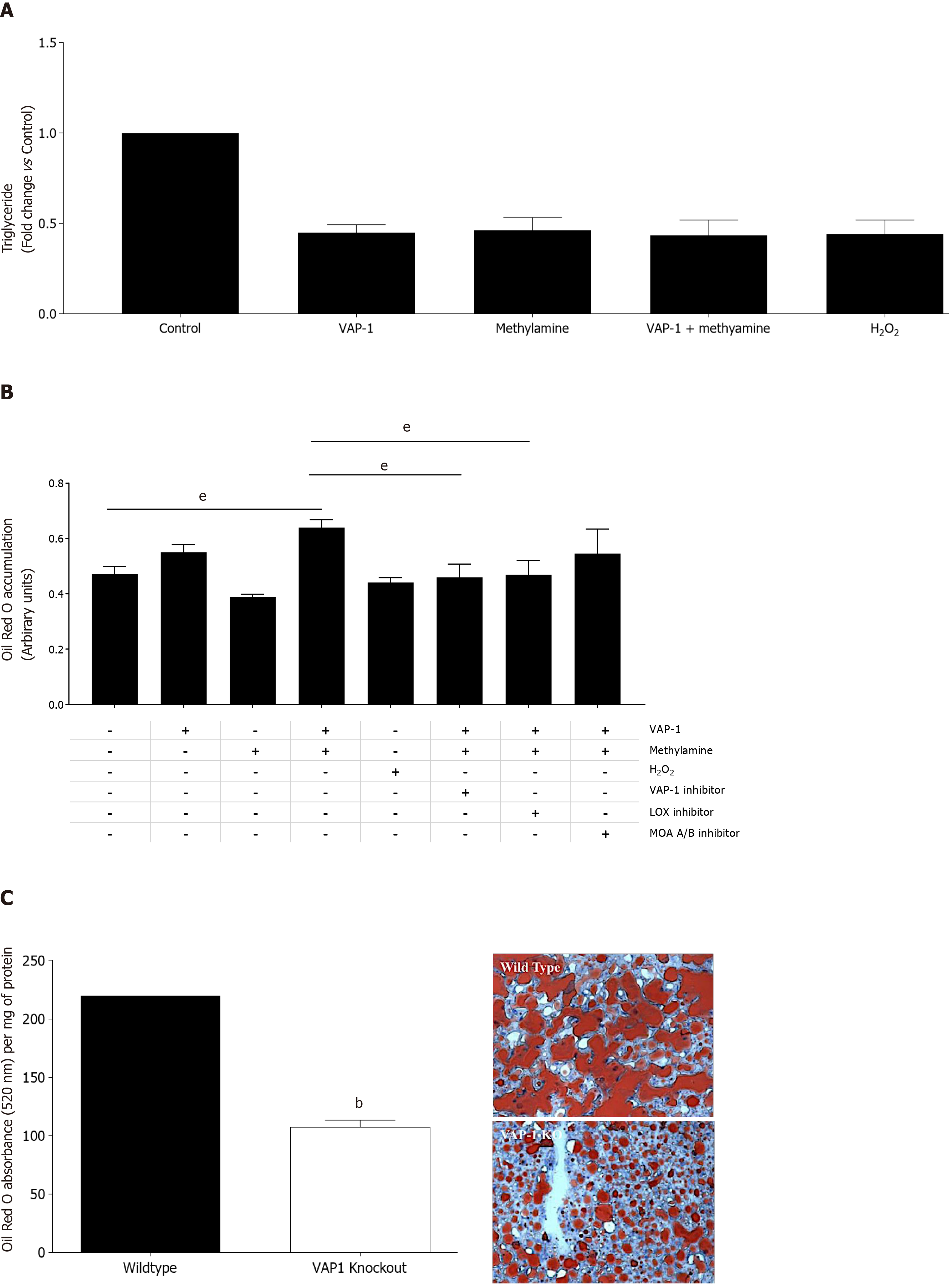
Figure 3 Activation of vascular adhesion protein-1 enzyme activity results in reduced triglyceride export and increased steatosis in human liver tissue and VAP-1/AOC3 knockout protects against high fat diet induced steatosis in mice.
A: PCLS were pretreated with either, methylamine 200 μm, vascular adhesion protein-1 (VAP-1) 500 ng, H2O2 10 μmol/L, or a combination of methylamine + VAP-1, benzylamine+VAP-1 for approximately 18 h and then 6 h with 250 μm fatty acid. Supernatants were collected after treatments and triglyceride secretion was quantified using a commercial assay (Cayman Chemical Company) according to manufacturer’s instructions. Data are triplicate samples from n = 2 normal livers ± SEM. P < 0.01 for all using a one way ANOVA; B: Lipid uptake in PCLS from normal liver tissue pretreated with either methylamine 200 μm, and rVAP1 500 ng/mL alone or in combination, or, H2O2 10 μmol/L. Some slices were exposed to the combination of methylamine and VAP-1 plus selective enzyme inhibitors: Bromoethylamine (VAP-1 inhibitor 400 μm), β-aminopropionitrile (lysyl oxidase inhibitor, BAPN 250 μm) or the Monoamine oxidase A and B inhibitors Clorgylline and Pargylline (both at 200 μm). After approximately 18 h incubation, slices were exposed to 250 μm oleic acid for 6 h. PCLS were fixed and stained with Oil RedO, which was solubilized and signal normalized to per 500 mg of tissue. Data are mean of triplicate samples from n = 2 normal livers ± SEM. Significance expressed as eP < 0.001 in one way ANOVA with Tukeys correction for multiple comparisons; C: Left - Accumulation of lipid in WT and VAP-1 KO mice fed on a high fat diet for 12 wk. 7um cryosections from WT and VAP-1 KO mouse livers were stained with ORO, which was then solubilized and signal expressed relative to protein concentration for each group of mice, Data are mean ± SEM of three mice per group. Significance expressed as bP < 0.01 one way ANOVA. Right – representative brightfield microscopy images of Oil red O stained cryosections from WT and VAP-1 KO mice.
- Citation: Shepherd EL, Karim S, Newsome PN, Lalor PF. Inhibition of vascular adhesion protein-1 modifies hepatic steatosis in vitro and in vivo. World J Hepatol 2020; 12(11): 931-948
- URL: https://www.wjgnet.com/1948-5182/full/v12/i11/931.htm
- DOI: https://dx.doi.org/10.4254/wjh.v12.i11.931


