Copyright
©The Author(s) 2020.
World J Hepatol. Nov 27, 2020; 12(11): 931-948
Published online Nov 27, 2020. doi: 10.4254/wjh.v12.i11.931
Published online Nov 27, 2020. doi: 10.4254/wjh.v12.i11.931
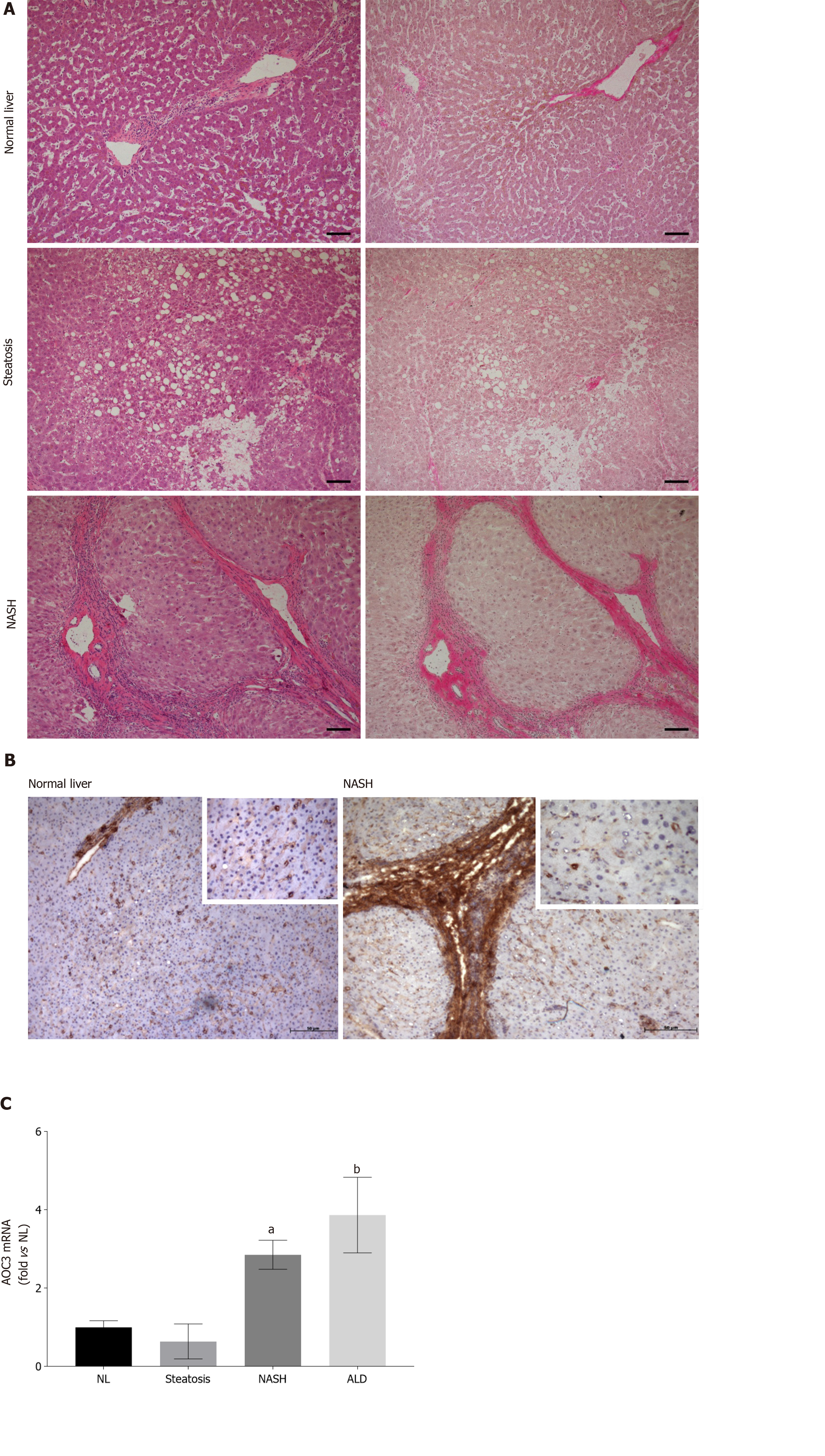
Figure 1 Hepatocellular expression of vascular adhesion protein-1 increases in nonalcoholic steatohepatitis.
A: Representative brightfield images of sections from indicated disease types stained with Hematoxylin and Eosin (left panels) and Van Geison’s Stain (right panels). Original magnification 10 ×, images representative of multiple fields of view from n = 3 Livers of each disease type. Scale bar is 100 µmol/L; B Immunohistochemical staining for vascular adhesion protein-1 (VAP-1) in representative acetone fixed frozen sections from normal and nonalcoholic steatohepatitis (NASH) livers. Isotype matched control antibody was negative (not shown). Fields were captured at 10 × original magnification with inset pictures captured at 40 × original magnification; C: Analysis of VAP-1 (AOC3) expression by quantitative qPCR analysis. mRNA expression of AOC3 in whole liver RNA from normal, steatotic, NASH, and alcohol-related cirrhosis (ALD) livers using fluidigm qPCR array®, run on triplicate arrays. Results are expressed as the mean fold change in gene expression normalized to pooled endogenous controls β-actin and GAPDH relative to normal livers defined as 1 ± SEM with means from five normal l, four steatotic, three NASH, and four ALD livers. aP < 0.05 or bP < 0.01 using a one way ANOVA with Bonferroni correction. NASH: Nonalcoholic steatohepatitis; ALD: Alcohol-related cirrhosis.
- Citation: Shepherd EL, Karim S, Newsome PN, Lalor PF. Inhibition of vascular adhesion protein-1 modifies hepatic steatosis in vitro and in vivo. World J Hepatol 2020; 12(11): 931-948
- URL: https://www.wjgnet.com/1948-5182/full/v12/i11/931.htm
- DOI: https://dx.doi.org/10.4254/wjh.v12.i11.931


