Copyright
©The Author(s) 2020.
World J Hepatol. Nov 27, 2020; 12(11): 1128-1135
Published online Nov 27, 2020. doi: 10.4254/wjh.v12.i11.1128
Published online Nov 27, 2020. doi: 10.4254/wjh.v12.i11.1128
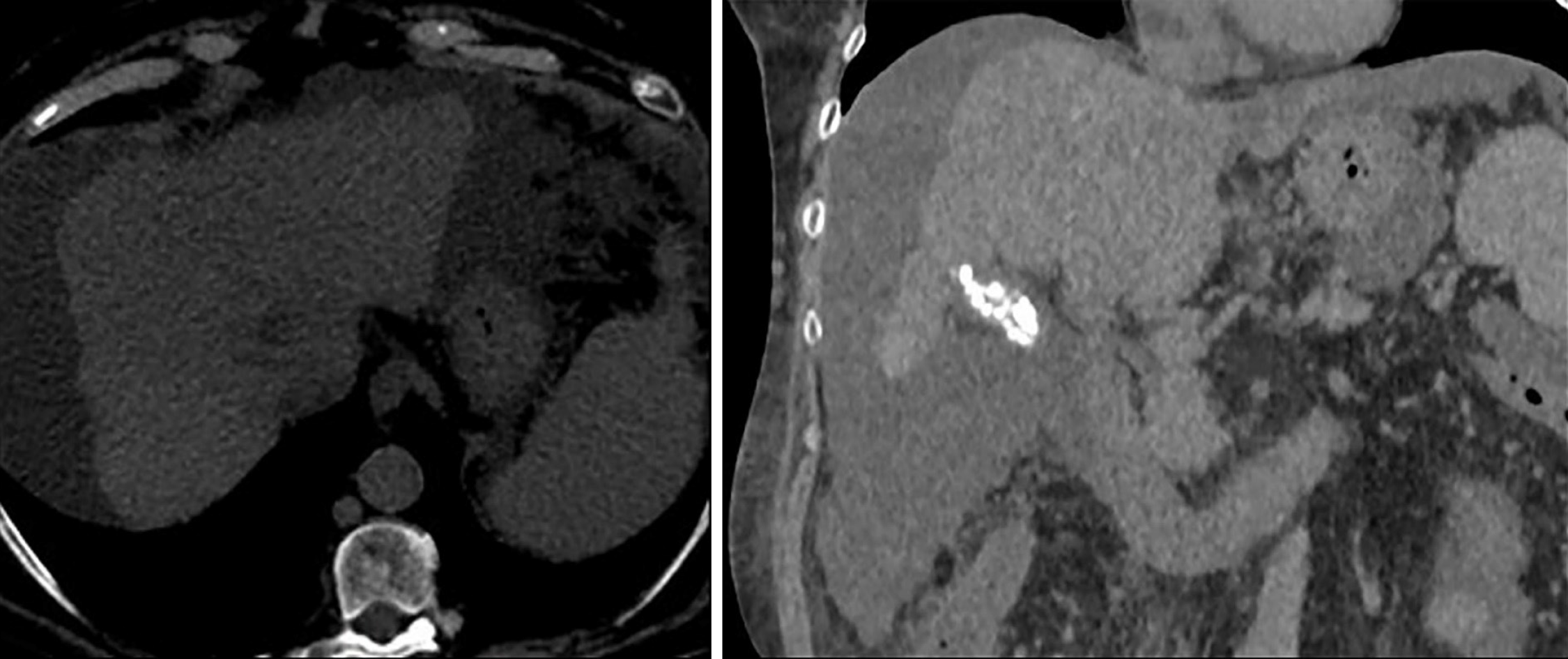
Figure 4 Non-contrast computed tomography scan axial view showing persistence of the heterogeneous lesion in the segment VIII seen previously, not delimitable in the current study.
Hypodensity persists in the inferior vena cava and right atrium suggestive of tumor extension, with apparent small reduction, although the intravascular tumor thrombus cannot be properly determined in a non-contrast computed tomography.
- Citation: Gomez-Puerto D, Mirallas O, Vidal-González J, Vargas V. Hepatocellular carcinoma with tumor thrombus extends to the right atrium and portal vein: A case report. World J Hepatol 2020; 12(11): 1128-1135
- URL: https://www.wjgnet.com/1948-5182/full/v12/i11/1128.htm
- DOI: https://dx.doi.org/10.4254/wjh.v12.i11.1128


